Venous Thromboembolism in Pregnancy
Siniša Franjić*
Independent Researcher, Europe
*Corresponding author: Siniša Franjić, Independent Researcher, Europe
Citation: Franjić S. Venous Thromboembolism in Pregnancy. Genesis J Gynaecol Obstet. 1(1):1-8.
Received: February 03, 2024 | Published: March 13, 2025
Copyright© 2025 genesis pub by Franjić S. CC BY-NC-ND 4.0 DEED. This is an open-access article distributedunder the terms of the Creative Commons Attribution-NonCommercial-No Derivatives 4.0 International License.,This allows others distribute, remix, tweak, and build upon the work, even commercially, as long as they credit the authors for the original creation.
Abstract
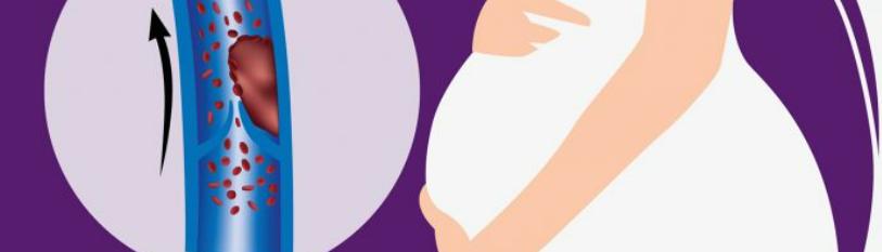
Swelling and sudden extreme pain caused by the intrusion of circulation, changes in the skin and its warmth, deadness and loss of motion of the influenced portion of the body are a few of the conceivable side effects of a thrombus. In any case, the genuine threat lies in the truth that portion of the thrombus can partitioned from the rest of the clot and travel to the heart, lungs or brain. Blocking the blood stream and oxygen supply to any of these organs requires crisis restorative consideration. In spite of the fact that this condition is uncommon, and most frequently influences individuals of the elderly populace, the infection can happen at any age. Venous thrombosis happens during pregnancy due to hormonal changes, expanded blood coagulation, or its clotting, which secures the mother and child from bleeding from the uterus, and due to the increment in the volume of the uterus, which can press on other organs and veins and hence moderate down the stream of blood to the lower limits and its way back to the heart.
Keywords
Venous Thromboembolism; DVT; Risk Factors; Pregnancy; Health
Introduction
Complicating 1 in 1500 pregnancies, venous thromboembolism (VTE) is a driving cause of maternal dreariness and mortality [1]. Additionally, in spite of this apparently low predominance, pregnancy confers a about 6–10-fold expanded hazard of VTE in women of comparable childbearing age. In one huge review cohort ponder, 94 of 127 (74.8%) pregnant women with reported profound venous thrombosis (DVT) created their clot during the antepartum period, with half identified some time recently 15 weeks and fewer than 30% analyzed after 20 weeks. In differentiate, most cases of pulmonary embolism (PE) created during the postpartum period (23 of 38; 60.5%) and PE was emphatically related with cesarean conveyance. In any case, the per diem chance of VTE is around triple to eightfold higher in the puerperium than during an identical antepartum interim. Aspiratory embolism is a driving cause of maternal mortality in the United States, contributing to 9.6% of such deaths. An untreated DVT presents a 25% hazard of PE, with a mortality rate of around 15% if undetected and untreated. On the other hand, if a DVT is instantly analyzed and treated, the hazard of PE is less than 5% and the chance of maternal mortality is less than 1%.
The expanded hazard of pregnancy-associated VTE reflects local and systemic components that relieve the chance of hemorrhage during placentation and the third organize of labor. Appreciation of the thrombotic chance of pregnancy requests information of the modern frameworks of coagulation and fibrinolysis and their inhibitors.
Conditions
A thrombosis (blood clot inside a blood vessel) can cause an irritation of the blood vessel lining (thrombophlebitis), which in turn can lead to a thromboembolism (hindrance of a blood vessel by a blood clot carried by the circulation from the location of beginning) [2]. Thrombi can involve the shallow or profound veins in the legs or pelvis. Shallow venous thrombosis more often than not includes the saphenous venous framework and is restricted to the lower leg. Shallow thrombophlebitis may be caused by the utilize of the lithotomy position amid birth. Profound venous thrombosis can include profound veins from the foot to the calf, to the thighs, or pelvis. In both areas, thrombi can remove and move to the lungs, causing a pneumonic embolism.
The three most common thromboembolic conditions happening during the postpartum period are shallow venous thrombosis, profound venous thrombosis, and pneumonic embolism. In spite of the fact that thromboembolic clutters happen in less than 1% of all postpartum ladies, pneumonic embolus can be deadly if a clot discourages the lung circulation; hence, early recognizable proof and treatment are paramount.
DVT
Deep venous thrombosis (DVT) happens in somewhat <1% of pregnancies [3]. The pregnant state increments the chance fivefold due to the venous stasis with the huge gravid uterus squeezing on the vena cava and the hypercoagulable state due to the increment in clotting variables. Cesarean conveyance encourage increments the hazard of DVT. In spite of the fact that clots including the shallow venous framework posture for all intents and purposes no peril and may be treated with absense of pain, DVT is related with pneumonic embolism in 40% of untreated cases. The hazard of death is expanded ten times when pneumonic embolism is unrecognized and untreated. Hence, early diagnosis and anticoagulation treatment are crucial.
Signs and indications of DVT incorporate profound leg torment, straight lines palpated along the calf, and delicacy and swelling of the lower limit. A 2-cm contrast in leg circumferences is also exceedingly suggestive. Tragically, none of these discoveries are exceptionally particular for DVT, and in truth, the examination is typical in half of patients with DVT. Subsequently, imaging tests are fundamental for confirmation. In pregnancy, the demonstrative test of choice is Doppler ultrasound imaging, which more often than not utilizes a 5- to 7.5-MHz Doppler transducer to degree venous blood stream with and without compression of the profound veins. This methodology is about as delicate and particular as the time-honored strategy of differentiate venography.
Management of DVT is essentially anticoagulation with bed rest and limit height. Anticoagulation treatment is the same as aspiratory embolism treatment with full intravenous dosages for 5 to 7 days, taken after by subcutaneous treatment for at slightest 3 months after the intense occasion. After 3 months, either full or prophylactic heparin measurements can be utilized for the leftover portion of the pregnancy and for 6 weeks postpartum. Patients who have extra chance variables for thromboembolism exterior of pregnancy may require long-term anticoagulation.
Platelet
Vasoconstriction and platelet accumulation are the introductory limitations on hemorrhage taking after vascular disturbance, especially in supply routes [1]. Vasoconstriction limits add up to blood stream to advance platelet plug arrangement. It also limits the measure of the plug required to deter blood stream through the vascular imperfection. Platelet attachment is at first intervened through von Willebrand factor (vWF), which is synthesized by megakaryocytes and endothelial cells. It is constitutively emitted but the most dynamic shapes are put away in endothelial Weibel-Palade bodies or platelet α-granules where the particle is discharged upon actuation. Upon vascular divider disturbance and after authoritative to subendothelial collagen, vWF experiences a conformational alter. This alter grants it to moreover tie to the platelet glycoprotein (GP) Ib/IX/V receptor to build up a hemostatic bridge between collagen in the harmed vessel divider and the platelet. The resultant GPIb/IX/V-vWF-collagen interaction encourages platelet attachment in the high-flow (shear stretch) state initiated by vasoconstriction. A moment platelet–collagen interaction is interceded by the official of collagen I and IV to the platelet GPIa/IIa (integrin α2β1) receptor in the low-flow settings made by the growing platelet plug.
Following collagen-mediated connection, a moment platelet receptor, GPIIb/IIIa (integrin α-IIb/β-3), is presently situated to serve as an elective extracellular lattice following location on platelet cell layers, allowing connection to subendothelial laminin, thrombospondin, fibronectin, vitronectin, and conceivably also vWF. In addition, GPIIb/IIIa-mediated platelet attachment at that point triggers calcium-dependent protein kinase C (PKC) actuation that actuates thromboxane A2 (TXA2) blend and platelet granule discharge. Next to vWF, platelet α-granules also contain different other clotting variables whereas densegranules contain adenosine diphosphate (ADP) and serotonin, which combine with TXA2 to potentiate vasoconstriction and advance advance platelet actuation. Platelets can moreover be actuated by thrombin, epinephrine, arachidonic corrosive and platelet-activating calculates. Platelet enactment, in turn, discharges more platelet surface GPIIb/ IIIa receptors that advance conglomeration by shaping interplatelet fibrinogen, fibronectin, and vitronectin bridges. Platelet accumulation in the setting of intaglio endothelium is avoided by dynamic blood stream and prostacyclin, nitric oxide, and ADPase.
During the platelet enactment prepare, anionic (adversely charged) phospholipids are exteriorized on the cell layer, making a perfect clotting surface. Platelet and endothelial cell actuation moreover actuates discharge of P-selectins from platelet α-granules and endothelial Weibel-Palade bodies that insert in the cell dividers to assist advance platelet and upgrade monocyte adherence. P-selectin authoritative too actuates expression of the strong procoagulant, tissue factor (TF), on monocytes to advance upgrade clotting (talked about afterward). These steps emphasize the basic part played by platelets, monocytes, and endothelial cells in advancing the classic coagulation cascade.
Risk Factors
Risk variables for thrombosis not special to pregnancy incorporate age over 35 years, obesity (body mass record ≥30kg/m2), paralysis, infection, smoking, nephrotic disorder, hyperviscosity disorders, malignancies, trauma, surgery, orthopedic strategies, and an earlier history of VTE [1]. There are also extreme pregnancy-specific nonreversible and reversible hazard variables that increment VTE chance. The nonreversible risk-factors incorporate expanded equality, numerous developments, preeclampsia, postpartum endomyometritis, postpartum hemorrhage requiring transfusion, agent vaginal conveyance and cesarean conveyance. New cesarean conveyance increments the chance of VTE indeed encourage than the fourfold increment in VTE chance related with nonemergent cesarean conveyances. Pregnancy-associated reversible chance variables incorporate ovarian hyperstimulation disorder, fixed status, long separate travel (>4–6 hours), hyperemesis related with drying out, and clinic remain. The chance for VTE endures for up to 28 days after a healing center remain amid pregnancy.
All three components of Virchow’s group of three – vascular stasis, hypercoagulability, and vascular damage – are show in pregnancy. Stasis comes about from increments in profound vein capacitance auxiliary to expanded circulating levels of estrogen and endothelial generation of prostacyclin and nitric oxide, coupled with compression of the second rate vena cava and pelvic veins by the broadening uterus. Interests, over 85% of DVT cases in pregnancy are left-sided, likely due to compression of the cleared out iliac vein by the right iliac course and the gravid uterus.
Imaging
Intravenous differentiate venography is the gold standard for DVT determination but is no longer utilized for the diagnosis of DVT in pregnancy since it has no more prominent symptomatic viability than VUS (venous ultrasonography) and is related with calculable radiation presentation [1]. Compression venous ultrasonography with or without color Doppler has developed as the favored introductory imaging methodology. It requires sonographic imaging of the common femoral vein at the inguinal tendon, and at that point appraisal of the other major venous frameworks of the leg, counting the more noteworthy saphenous, the shallow femoral, and the popliteal veins to the profound veins of the calf. Pressure is connected to the transducer to decide the compressibility of the vein lumen beneath duplex and color stream Doppler imaging. Noncompressibility of the venous lumen in a transverse arrange with tender pressure from the ultrasound test utilizing duplex and color stream Doppler imaging would be demonstrative for a venous thrombosis. The by and large affectability and specificity of VUS approach 100% for proximal vein thromboses with somewhat less viability for identifying separated calf vein DVT (affectability 92.5%, specificity 98.7%, and precision 97.2%).
A meta-analysis of 14 thinks about comparing the viability of attractive reverberation imaging (MRI) venography with a reference standard in nonpregnant patients with suspected DVT appeared a pooled affectability of 91.5% and a pooled specificity of 94.8%, with a higher affectability for proximal than distal DVT. In this way, MRI has proportionate affectability and specificity to VUS for the diagnosis of DVT. It may, be that as it may, be valuable for recognizing iliofemoral/pelvic vein thromboses which are ineffectively visualized by VUS and account for 11% of DVT in pregnancy, compared with fair 1% in the nonpregnant state.
Hormonal Contraception
Confirmatory tests for VTE incorporate D-dimer testing, CTPA, Ventilation/Perfusion (V/Q) checks, and lower limit Doppler testing [4]. Utilize of D-dimer testing ought to be confined to barring VTE in patients with a low pretest likelihood. D-dimer testing was not performed for this understanding as she was as of now at tall likelihood for PE. CTPA is the demonstrative methodology of choice with affectability and specificity extending from 83 to 100% and 86 to 100% individually. V/Q looks are secondline imaging thinks about, ordinarily utilized for patients with a contraindication to CTPA such as hypersensitivity to differentiate, kidney failure, or failure to get intravenous get to. Aspiratory angiography was once considered the gold standard to analyze PE, but is once in a while utilized nowadays due to the invasiveness of the test and the accessibility of CTPA. Lower limit Doppler thinks about can play a part in the assessment of VTE. A persistent with a positive Doppler ponder for profound vein thrombosis (DVT) and aspiratory side effects can be assumed to have PE and can be overseen in like manner. In any case, a negative think about does not run the show out PE. Here, CTPA affirmed the nearness of PE.
Usual treatment for VTE incorporates anticoagulation with unfractionated or low-molecular-weight heparin and warfarin. Thrombolytics may be considered in patients with broad DVTs including the iliac and femoral veins as well as in patients with enormous PEs. Inferior vena cava (IVC) channels may be put in patients with contraindications to therapeutic administration or in patients with repetitive DVTs. As this persistent was hemodynamically steady, she was treated with low-molecular-weight heparin whereas starting warfarin to helpful dosages. An IVC channel was not demonstrated given this was her to begin with VTE event.
Once VTE is affirmed, encourage assessment for fundamental coagulopathy may be performed. As a few parts of the research facility assessment are modified by anticoagulation, research facility considers ought to be drawn earlier to start of such treatment. The assessment regularly incorporates Protein S and C lacks, figure V Leiden transformation, antiphospholipid counter acting agent, antithrombin III lack, prothrombin quality transformations, and lupus anticoagulant. The nearness of an fundamental hypercoagulable state may require long-term anticoagulation. Schedule screening for these disorders is not suggested earlier to beginning combined hormonal contraception due to the irregularity of the conditions and the tall taken a toll of screening. A thrombophilia workup was requested and found negative in this quiet. As the VTE likely stemmed from her combined verbal prophylactic utilize in conjunction with her corpulence and later stability, a three-month treatment course of anticoagulation was prescribed. She was counseled on modifiable chance variables counting weight diminishment and the require to dodge drawn out immobility.
Combined hormonal contraceptives are contraindicated in patients with a history of a VTE. Be that as it may, due to the by and large expanded chance of VTE during pregnancy, successful contraception is prompted in this quiet. In spite of the fact that combined hormonal prophylactic utilize is related with up to a fourfold increment in chance of VTE in a nonpregnant female, that supreme hazard of VTE is still lower than women in the pregnant and puerperium state. As in other patients, long-acting reversible contraception (LARC) strategies are favored. Suggestions for prophylactic administration in a persistent with VTE are laid out by the Centers for Disease Control and Prevention (CDC) in their “US medical eligibility criteria for contraceptive use, 2010”. These rules suggest the cessation of combined hormonal contraceptives in any quiet with intense VTE or history of VTE in any case of current anticoagulation treatment or inclining components. In patients with DVT or PE, progestin-only prophylactic pills, station medroxyprogesterone acetic acid derivation infusions, etonogestrel inserts, and levonorgestrel or copper IUDs are all worthy choices, and are category 2 (preferences of the strategy by and large exceed the hypothetical or demonstrated dangers). This quiet was teaching to cease combined verbal contraceptives and she chose to start the levonorgestrel IUD (intrauterine gadget), which may too offer assistance avoid overwhelming menstrual dying whereas the quiet is anticoagulated.
PT
Anatomically, the liver is for the most part not discernable underneath the right costal edge auxiliary to the broadening gravid uterus [5]. Both liver measure and net appearance do not alter during pregnancy. Moreover, negligible histologic changes happen in pregnancy. These incorporate cellular and atomic pleomorphism, expanded numbers of binucleate cells, steatosis, expanded cellular and atomic glycogen, hypertrophied Kupffer cells, and a few lymphocytic invasions of the entry tracts. During typical pregnancy, plasma volume is expanded by 50% to 70% and comes to its greatest by 32 to 34 weeks of pregnancy. This comes about in hemodilution of all serum protein concentrations. Serum egg whites concentration diminishes, but the rates of egg whites blend and catabolism are not influenced. Hypoalbuminemia can happen in the third trimester notwithstanding of liver disease. Be that as it may, prolongation of prothrombin time (PT) and hyperbilirubinemia would raise the concern of critical liver disease. Other serum protein concentrations such as alpha-2-macroglobulin, alpha-1antitrypsin, and ceruloplasmin are expanded. Pregnancy is characterized by a hypercoagulable state and expanded dangers of profound venous thrombosis. Nearly all coagulation components increment amid pregnancy but protein S. Additionally, fibrinolysis is hindered.
This physiologic state tends to diminish the hazard of bleeding during conveyance; in any case, it increments the hazard of maternal venous thrombosis. In fact, PT is for the most part not influenced during pregnancy and any changes in PT ought to be considered unusual and legitimize encourage examination. Serum lipid concentrations increment in late pregnancy auxiliary to expanded levels of estrogen and progesterone. Rises in estrogen and progesterone levels improve cholestasis and generation of lithogenic bile and diminish gallbladder motility. These changes incline pregnant women for gallstone and biliary slime arrangement. Estimation of serum lipids in pregnancy is seldom valuable unless in the setting of intense pancreatitis. During pregnancy, the add up to red cell mass is really expanded but quickly diminishes as pregnancy ceases. The cardiac yield increments but the supreme hepatic blood stream remains unaltered. Plasma volume increments extraordinarily during the moment trimester coming about in falling hemoglobin levels due to hemodilution. Serum aminotransferases [alanine aminotransferase (ALT) and aspartate aminotransferase (AST)] are not modified by pregnancy and are considered the most valuable test demonstrative of hepatocellular damage. Serum soluble phosphatase action levels rise during the third trimester as a result of expanded generation of placental and bone isoenzymes. In fact, estimation of serum soluble phosphatase action is not a appropriate test for the diagnosis of cholestasis during pregnancy. Add up to and free bilirubin concentrations are diminished during pregnancy somewhat due to hemodilution.
Gilbert’s disorder is characterized by an increment in unconjugated hyperbilirubinemia with outright ordinary aminotransferases. The administration of pregnant women with Gilbert’s syndrome is not any diverse than those who are not pregnant, and therapeutic consolation ought to be given. The disorder of hemolysis, elevated liver enzymes, and low platelets (HELLP) is related with circuitous hyperbilirubinemia and ought to be separated from Gilbert’s disorder. Pregnancy is also related with mellow increment in serum bile acids auxiliary to diminished bile salt transport. Estimation of serum bile acid concentrations may be valuable for the determination of cholestasis during pregnancy.
Treatment
Once there is a tall clinical doubt of DVT or PE, treatment ought to be commenced unless there is a solid contraindication to anticoagulant treatment [6]. Security of the mother and the fetus must be taken into thought when making choices on treatment. LMWH (low molecular weight heparin) has a more great security profile than UFH (Unfractionated heparin), more helpful once or twice every day dosing, and nonappearance of the require for standard research facility checking with the activated partial thromboplastin time (APTT) which is less dependable in pregnancy. Be that as it may, checking utilizing anti-Xa movement may be considered in people with extremes of body weight (weight <50 kg or >90 kg).
The weight balanced twice every day dosing regimen of LMWH is favored over the once day by day dosing in arrange to compensate for the increment glomerular filtration rate that happens in the moment trimester of pregnancy.
The rate of heparin-induced thrombocytopenia is low taking after LMWH utilize in pregnancy; unless there is a history of past HIT (heparin-induced thrombocytopaenia) from UFH, platelet number checking is not necessary.
Conclusion
Venous thrombosis is considered to be the prepare of intravital intravascular coagulation of blood in the veins. Hazard variables for its event are: fixed status, postoperative condition, dangerous tumors, hormone treatment, injury and pregnancy. The most common localizations of thrombus are the femoral vein, lower leg veins, popliteal and subclavian veins. The rate of thromboembolism in the prenatal period is 0.2%, and triples after conveyance. Cesarean area increments the frequency to almost 1-2%. Aspiratory embolism, with a mortality of around 15%, happens in around 50% of patients with affirmed profound vein thrombosis. Early determination and satisfactory restorative strategies definitely diminish the frequency of aspiratory embolism and death.
References
Genesis Scientific Publication is licensed under CC BY-NC-ND 4.0![]()
![]()
![]()
![]()
