Uterine Intravascular Leiomyomatosis Extending to the Inferior Vena Cava and Right Heart Chamber
Boukhili Houssem1*, Saoudi Wassim1, Ben Abdesslem Aymen1, Ernez Hajri Samia1, Cherif Taieb2, Imen Mgarech2 and Kaabia Ons3
1University of Sousse Faculty de Medicine of Sousse, Farhat Hached Hospital of Sousse Cardiology Department /LR12SP09
2Department of cardiovascular and thoracic surgery, Sahloul Hospital of Sousse, Tunisia
3Department of Gynecology, University of Sousse Faculty de Medicine of Sousse, Farhat Hached Hospital of Sousse
*Corresponding author: Boukhili Houssem, Faculty of medicine Rue Mohamed Karoui Sousse, Tunisia.
Citation: Houseem B, Wassim S, Aymen BA, Samia EH, Taieb C, Mgarech I, Ons K. Uterine Intravascular Leiomyomatosis Extending to the Inferior Vena Cava and Right Heart Chambers Genesis J Surg Med. 1(1):1-8.
Received: June 20, 2022 | Published: July 07, 2022
Copyright© 2022 genesis pub by Houssem B. CC BY-NC-ND 4.0 DEED. This is an open-access article distributed under the terms of the Creative Commons Attribution-Non Commercial-No Derivatives 4.0 International License. This allows others distribute, remix, tweak, and build upon the work, even commercially, as long as they credit the authors for the original creation.
DOI: https://doi.org/10.52793/GJSM.2022.1(1)-3
Abstract
Background: Uterine leiomyoma is a benign tumor that occurs in young women of reproductive and pre- menopausal age. Intravascular leiomyomatosis is a very rare condition and cardiac extension involving the inferior vena cava and right atrium is even rarer. We report a case of intravascular leiomyomatosis extending through the inferior vena cava and right heart chambers.
Case summary: A 43-year-old woman with a free past medical history was referred to our department to take care of abdominal pain and dyspnea. A transthoracic echocardiography was performed showing an intracardiac mass in the right atrium and the inferior vena cava. A thoracic-abdominal-pelvic computed tomography angiography and an abdominopelvic MRI were performed suspecting the diagnosis of intravenous leiomyomatosis extending from the ovarian vein to the inferior vena cava and the right atrium. The patient was referred to surgery and a total hysterectomy with bilateral adnexectomy associated with a delivery of the tumor through the inferior vena cava was performed with a confirmation of the diagnosis in the histological examination.
Conclusion: Intravenous leiomyomatosis is a rare condition with a challenging diagnosis due to its atypical clinical presentation and insidious disease progression. The extension to cardiac cavities presents an evolutionary turning point with a poor prognosis needing a total resection of the tumor.
Keywords
Leiomyomatosis; Intravascular and intracardiac extension; Cardiac mass; Echocardiography
Introduction
Uterine leiomyoma is a benign tumor that occurs in young women of reproductive and pre- menopausal age. Extra-uterine expression is uncommon and presents in four entities: intravascular leiomyomatosis (IVL), disseminated peritoneal leiomyomatosis (DPL), benign metastasizing leiomyoma (BML), and hereditary leiomyomatosis.
Intravascular leiomyomatosis is a very rare condition and cardiac extension involving the inferior vena cava and right atrium is even rarer occurring in approximately 10 to 40% of cases [1,2]. Knowledge of this pathology is important as it requires special and multidisciplinary management depending on the degree of extension.
We report a case of intravascular leiomyomatosis extending to the inferior vena cava and right heart chambers and we discuss through a review of the literature the clinical presentation, the diagnosis modalities, the therapeutic management, and the evolution of this variety of tumors.
Patient and Observation
Patient information
This concerns the case of a 43-year-old woman with no notable medical or surgical history, G5P2A3, with three medical interruptions of pregnancy, who presented with abdominal pain associated with dyspnea and heaviness of the lower limbs evolving for six months.
Clinical findings
A complete clinical and gynecological examination showed no anomalies except a sinus tachycardia on the electrocardiogram at rest.
Diagnostic assessment
The patient underwent an abdominal-pelvic ultrasound which revealed a dilated right ovarian vein with an extensive hyperechoic content extending to the supra-renal inferior vena cava and the right atrium. A trans-thoracic and trans-esophageal echocardiogram was performed and revealed the presence of a mobile echogenic mass, with a serpentine appearance in the inferior vena cava and the right atrium measuring 70 * 24 mm. The mass was not adherent to the wall of the right atrium and passed on either side of the tricuspid valve without causing obstruction or functional impairment of the right cavity. (Figure 1; Figure 2). We discussed the differential diagnoses for cardiac mass that include a myxoma which presents the most frequent type of benign cardiac tumor. We excluded this diagnosis because of the echocardiography characteristics and the origin of the tumor. A thrombus, metastatic cancer but we hadn’t favorable context, and lipoma were also considered.

Figure 1: Subcostal echocardiographic section showing a hyperechoic mass in the inferior vena cava extending to the right atrium.

Figure 2: Four-chamber echocardiographic section showing the presence of a well-defined hyperechoic mass in the right atrium.
Further investigation was initiated including tumor marker testing (ACE, CA 19-9, CA 125) which came back negative; immunological screening (anti-nuclear antibodies, anti-SSA, anti- SSB, anti-cardiolipin antibodies, anti-phospholipid antibodies) and thrombophilia screening (Protein C, protein S, antithrombin III and factor V mutation testing) were normal.
The patient underwent a thoracic-abdominal-pelvic computed tomography angiography that showed a hypervascular tumor originating from the pelvic region from the right ovarian vena extending to the inferior vena cava and the right atrium. The diagnosis of intravenous leiomyomatosis extending from the ovarian vein to the inferior vena cava and the right atrium was suspected (Figure 3).

Figure 3: Longitudinal section of thoracic-abdominal-pelvic CT scan showing the tumor mass extending from the right utero-ovarian vein to the right atrium.
We completed with an abdominal-pelvic MRI which demonstrated the presence of a hypervascular lesion in the right ovarian vein that extended from a 5 cm lesion in the right lateral uterine wall, enhancing with gadolinium in a manner identical to the myometrium and extending to the inferior vena cava with significant cardiac extension, suggestive of intravenous leiomyomatosis (Figure 4).


Figure 4: 3D reconstruction of abdominopelvic MRI showing an extensive intravascular leiomyomatosis extending up to the right atrium.
Therapeutic Interventions
The therapeutic management was discussed by a multidisciplinary team including cardiologists, cardiovascular surgeons, and gynecologists and we decided to perform a total hysterectomy with bilateral adnexectomy associated with a delivery of the tumor through the inferior vena cava under extracorporeal circulation. Our patient underwent surgery on 31/03/2023 (Figure 5, 6).

Figure 5: Extraction of the tumor mass under extracorporeal circulation.

Figure 6: Operative specimen of total hysterectomy and bilateral annexectomy.
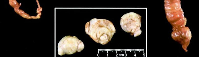
The gross examination of the surgical specimen showed multiple grey-whitish firm fibroids including the right lateral uterine wall and the right broad ligament. These tumors ranged in size from 5 mm to 5 cm with a fasciculate aspect on the cut surface. These tumors spread into fallopian tube vessels and utero-ovarian veins forming an oblong mass of 26 cm in length into the vena cava (Figure 7).

Figure 7 (A): Gross examination of total hysterectomy with bilateral salpingo-oophorectomy specimen with inferior vena cava (arrow) and thrombotic mass (arrowhead).

Figure 7 (B): On the cut surface, multiple fasciculate and grey-whitish nodules involving the right uterine wall.
Microscopically, the tumors consisted of intravascular nodules of intersecting fascicles of spindle-shaped cells originating from venous walls. The neoplastic cells have indistinct borders, eosinophilic cytoplasm, and centrally located and blunt-ended cigar- shaped nuclei without atypia. Few mitotic figures were seen. The mitotic count was 1 per 10 high-power fields. The tumor showed an increased vasculature and foci of hyalinization and hemorrhage. In an immunohistochemical study, the neoplastic cells were diffusely positive for desmin, smooth muscle antigen (SMA), estrogen, and progesterone receptors (Figure 8). Therefore, the diagnosis of uterine intravascular leiomyomatosis (UIVL) was made.

Figure 8: Photomicrographs of uterine intravascular leiomyomatosis showing A. intravenous development (Hematoxylin and eosin, HE x40) B. interlacing fascicle of spindle-shaped cells with foci of hyalinization (HE x200) C, D, E, and F. Immunohistochemistry study showed positivity for (C) desmin (magnification x100) (D) smooth muscle actin (magnification x400) (E) estrogen receptor (magnification x200) (F) progesterone receptor (magnification x100).
Discussion
The diagnosis of intravascular leiomyomatosis is challenging due to its rarity, atypical clinical presentation, and often incidental discovery.
Trans-thoracic echocardiography is of great importance in cases with extension to the right heart chambers, as it can suggest the diagnosis when a mobile, echogenic mass originating from the inferior vena cava and protruding into the right atrium is identified, with prognostic implications due to the risk of arrhythmia, syncope, right heart failure, or even death from complete tricuspid annular obstruction [3,4].
Imaging is crucial for better characterization of this pathology and assessing its extent, particularly with thoracic-abdominopelvic computed tomography (CT) and abdominal-pelvic magnetic resonance imaging (MRI) to evaluate the benign tumor's extension, which guides therapeutic strategy based on location and extent [5].
In the literature, preoperative classification is based on the degree of vascular extension according to imaging findings [8]. Definitive diagnosis relies on histopathological examination of the surgical specimen, which typically shows smooth muscle cells resembling those present in the uterus, with positive staining for actin, desmin, and hormonal receptors for progesterone and estrogen [10]. In our case, the diagnosis is intravascular leiomyomatosis stage III, necessitating total hysterectomy and bilateral adnexectomy due to the hormonal sensitivity of the tumor, along with intravascular and cardiac tumor resection.
The main risk of intravascular leiomyomatosis is recurrence and it represents up to 30% that’s why a regular follow-up involving the use of imaging techniques is essential to detect any potential recurrence [10].
Follow-up and Outcomes
The immediate postoperative evolution was favorable. The patient does not present any functional complaints, and it has been decided to follow the patient according to the literature data, with a follow-up plan as follows: We will complete a thoracic-abdominal-pelvic CT scan between the 3rd and 6th month post-operative, and then every 2 to 5 years as per the literature data [9].
Conclusion
Intravenous leiomyomatosis is a rare condition with a challenging diagnosis due to its atypical clinical presentation and insidious disease progression. The use of multiple imaging modalities is crucial for guiding multidisciplinary therapeutic management. Definitive diagnosis relies on histopathological examination, and treatment is primarily surgical. Regular follow-up is important to monitor for recurrence.
Learning Objectives
- Cardiac mass has multiple etiologies and it requires a range of complementary examinations to determine the etiology
- The use of multiple imaging modalities for diagnosis, tumor extension, therapeutic decision, and follow-up is crucial.
- Therapeutic management needs the coordination of the heart team and a multidisciplinary team.
Disclosures
The authors have nothing to disclose.
Funding
There was no funding.
Ethical Approval
The case report was approved by the ethics committee of Hospital Farhat Hached of Sousse, Tunisia.
Consent
Written informed consent was obtained from the patient.
References
Genesis Scientific Publication is licensed under CC BY-NC-ND 4.0![]()
![]()
![]()
![]()
