TMD A Modern Lifestyle Disease-Correlation Between Cranio-Cervical Posture and Presence of TMD Using Lateral Cephalogram as A Diagnostic Tool
Faisal Taiyebali Zardi1, Brajesh Gupta2, Rishika Reddy3*, Nagalaxmi V4, Srishitha Enaganti Rao5 and Sunayana Abbagalla6
Abstract
Introduction: Temporomandibular disease (TMD) etiology is multifactorial and is related to many perpetuating, predisposing and initiating factors. TMD is a range of conditions commonly characterized by heterogenous signs and symptoms and is reported to be one of the most common musculoskeletal/neuromuscular disorders and is often correlated to headache. The stomatognathic system requires a stable muscular – skeletal posture to perform its multiple daily functions. Modern habits like excessive use of cellphones, computers and sedentary lifestyle can be the cause for TMD.
Aim: To investigate the correlation between the cranio-cervical posture (CCP) and TMDs evaluated by cephalometric analysis.
Method: 100 individuals aged between 20 to 40 years were evaluated by cephalometric methods. From cephalometric analysis, cranio-cervical angle is evaluated to know the posture. A proper case history and TMJ palpation was done to know the temporomandibular joint clicks and tenderness to co – relate it with cephalometric analysis.
Results: In the present study the correlation between TMD and posture were evaluated using lateral cephalogram where the p-value was 0.004 which was considered statistically significant.
Conclusion: The present study concludes that improper CCP can cause TMD in patients.
Keywords
Cranio-cervical posture; Temporomandibular disorders; Lateral cephalogram; Mc Gregor plane
Abbreviations
Introduction
Temporomandibular disorders (TMDs), with a reporting prevalence between 28% and 88%, are common in young people aged 20 to 40 years and are most common in females [1]. TMD is a comprehensive disease involving joints, muscles, and nerves, which is mainly characterized by the restriction, deviation, deflection of mandibular movement, clicking or popping sound of temporomandibular joint (TMJ), pain in masticatory muscles, anterior ear, and the TMJ region [2,4].
The aetiology of TMD can be a variety of factors, including trauma, stress, mandibular dysfunction, malocclusion and cranio-cervical posture [5]. Studies found that TMD patients had a higher cervical anteversion and an increased cranio-cervical angle [6]. Posture can be considered as an etiologic factor, since studies regarding to patterns of mandibular, head and neck movements have been demonstrating biomechanical associations between temporomandibular and cervical spine systems.
The upper cervical spine is formed by Atlanta- occipital joint and the Atlanta- axial joint [7]. There are two types of spinal curve in the human body viz. Primary curve & the Secondary curve. A human baby is born with primary curve known as kyphotic curve [8]. Later, cervical portion is affected by various factors like craniofacial, morphology, orthodontic therapy, lifestyle habits, occupation etc.
The Forward Head Position (FHD) is characterized by an extension of the head together with the upper cervical spine (C1 to C3), accompanied by a flexion of the lower cervical spine (C4 to C7), whereby the cervical curvature is increased, a condition called hyper lordosis [9]. The hyperextension of upper cervical and a straightening of the lower cervical is common in TMD patients. The muscular activity resulting from cranio cervical extension of the head produces an elevation and retrusion force that acts on the mandible, which results in decrease of the physiological free-way space of TMJ [10]. On the other hand, the tissue elasticity, mainly represented by visco-elastic properties of the muscular and tendinous connective tissues, can also influence the mandibular postural position, when they’re stretched as a result of FHD (Figure 1).

Figure 1: Masticatory system functional alteration caused by Forward Head Position (FHD) [11].
Tension of the masticatory and suprahyoid muscles increases, which leads to mandibular elevation and retrusion. Due to the decrease in free-way space of TMJ, movement represented by the path of mandibular closure now occurs farther backwards than usual which makes the initial occlusal contacts locate further back than the maximal intercuspidal position (MIP) [11].
The aim of our study was to investigate the correlation between the cranio-cervical posture and TMDs by cephalometric analysis.
Methodology
Patient selection
100 patients between the age of 20 to 40 years, selected from the OPD of Oral Medicine and Radiology department in Sri Sai College of Dental Surgery, Vikarabad.
Patients’ exclusion criteria were – developmental abnormalities, skeletal abnormalities, trauma patients, history of head and neck surgery, post orthodontic treatment, pregnant women and lactating mother.
These 100 patients were evaluated by cephalometric analysis. For cephalometric analysis cranio-cervical angle was calculated using Mc Gregor plane to know the posture. A proper case history and TMJ palpation was performed to evaluate clicks and tenderness in the TMJ area.
An in-detail case history which consisted of personal history, family history, habit and para - functional habits, occupation and past medical history was recroded. Then patients were instructed to stand in their normal posture for lateral cephalogram exposure. After the exposure cephalometric analysis was performed to calculate cranio-cervical angle using Mc gregor plane with scale and sketch pen. All the patients TMJ was palpated by extra – articular and intra-articular methods (Figure 2).

Figure 2: Gergor plane on subject using ruler and sketch pen.
Palpation was done standing 12 o’clock position. Little finger was placed in external auditory meatus (during which posterior pole of condyle was palpated), with this method capsular tenderness was also palpated. Then for extra – articular examination index finger was placed on the pre - auricular region about 1.5 cm medial to the tragus of ear (lateral pole of condyle was palpated).
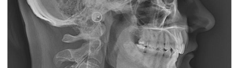
Mc gregor plane was calculated by drawing a plane that touches the base of the occipital bone to reach the posterior nasal spine and another plane from the apex of the odontoid process of C2 to the most anterior and inferior point of the body of C2 [12]. The angle between 96° to 106° were considered as normal head posture and angle below 96° were considered as patient having forward head posture as stated by Mariano Rocabado (1983) [20] (Figure 3).

Figure 3: Mc gregor plane on subject using ruler and sketch pen.
Results
In the present study 100 patients were evaluated to check relation between cervical posture and TMD.
A. Distribution of subjects on angle with symptomatic and with asymptomatic.
In the present study the minimum angle was 80° and maximum angle was 106°.
B. Distribution of symptomatic and asymptomatic subjects with forward head posture.
In the present study the distribution of symptomatic subjects with forward head position were 49, asymptomatic subjects with normal head posture were 7. Asymptomatic subjects with forward head posture were 26 and asymptomatic subjects with normal head posture were 18. The P- value was 0.042 which is statistically significant (Figure 4).

Figure 4: Distribution of subjects having TMD and no TMD.
The table (table 2) suggests a positive correlation (Pearson's r = 0.0042, p < 0.05) between ANGLE and TMD, based on a two-tailed significance test with a sample size of 100. This indicates a statistically significant but very weak relationship between the two variables.
Discussion
Temporomandibular disorders (TMD) are a collective term for a group of musculoskeletal conditions involving pain and/or dysfunction in the masticatory muscles, temporomandibular joints (TMJ), and associated structures [1]. Although TMD is defined by pain and dysfunction in the orofacial region; common painful and non-painful comorbidities of TMD include headaches, neck and back pain, fibromyalgia, irritable bowel syndrome, tinnitus, chronic fatigue syndrome, insomnia, depression, and anxiety [2].
The aetiology of temporomandibular disorder (TMD) is multifactorial. Posture being one of the reasons. One of the major reasons of forward head posture is modern lifestyle. Excessive use of computers, phones, long working hours, long travelling hours for work and education, slouched sitting. This posture creates altered length-tension relationships of the muscles attached to the jaw and can trigger hypertonus (increased tone or contraction) activity of some of those muscles. Imbalanced muscle forces around the jaw, including the suprahyoid muscles, are usually weaker compared to the masseter and temporalis, which close the jaw. The constant fight between muscles that perform depression and elevation of the mandible causes parafunction.
With forward head posture we increase the distance from chin to sternum stretching the hyoid muscles, which in turn will have the tendency to pull mandible back and down [13] (Figure 5).

Figure 5: Forward head posture13.
This could be considered as a “domino effect” i.e. the head moves forward shifting the centre of gravity to compensate that the upper body drifts backwards, to compensate for the upper body shift the hips tilt forward. So, the forward head position can be the cause for not only head/neck problems, but also mid-back and low- back problems [13].
The main finding of this study was that there was significant correlation of forward neck position with presence of temporomandibular disorder. Lateral cephalogram was selected to evaluate cranio - cervico- mandibular structures as it gives a more objective visualization without any influence of soft tissues [12].
The present study is in accordance with the study conducted by Priscilla Weber et al., (2012) they concluded saying cephalometric analysis seems to be the most appropriate since it provides a more objective perspective of the bone structures independent of the soft tissues [12].
In the current study the 49 out of 100 subjects having forward head posture had temporomandibular disorder showing in which nearly half of the subjects showing the correlation between forward head posture and TMDs, this is similar to the review article by Cuccia et al., who studied the relationship between the stomatognathic system and body posture and concluded that tension in the stomatognathic system can lead to impaired neural control of posture. If proprioceptive mechanism of the stomatognathic system is improper and inaccurate, then body position and head control may be affected [14].
Lateral cephalogram helped in this study to calculate the cranio-cervico-mandibular structures using Mc gregor plane which is similar to a study by Chu-Qiao Xiao er al., (2022) as they concluded saying TMD patients with TMJ pain showed increased FHP compared to other groups, and FHP became more significant as TMD severity increased in male patients, indicating the FHP may be a significant factor in the onset of TMJ discomfort. The aberrant head and neck posture of the patient may be taken into account in the clinical assessment of TMDs [15].
Basically, a change of head posture can produce a change of mandibular position. Several investigations have proven such a statement. Daily demonstrated that a bite opening experimentally produced by a mechanical device was accompanied by a significant cranio- cervical extension [16]. In patients who received immediate complete dentures, Tallgren, et al. determined that changes in the mandibular inclination due to reabsorption of the bone ridges were accompanied by changes in the craniocervical posture [17,18].
Middle age subjects having forward head posture is common because of excessive laptop and phone usage in unhealthy posture which is in accordance with a study conducted by Seong-Yeol Kim et al., to see the effect of duration of smartphone use on neck and shoulder muscle fatigue and pain was in adults with forward head posture. They concluded saying pain and fatigue worsened with longer smartphone use due to posture [21].
By this we can conclude temporomandibular joint disorder is a modern lifestyle disease associated with prolonged use of computers and mobile devices and sedentary behaviors which may contribute to poor posture. Poor posture can affect the biomechanics of the jaw and neck potentially contributing to TMDs. A sedentary lifestyle with limited physical activity can contribute to muscle imbalances and stiffness, potentially affecting the muscles around the jaw and causing TMDs.
Strength of the study
The present study evaluated the correlation between the cranio – cervical posture and TMD using cephalometric analysis. This study is first of its kind to correlate the forward head posture with TMD using lateral cephalogram. The study evaluated the posture using Mc gregor plane on lateral cephalogram. The results obtained in this study determined the presence of relation between forward head posture and temporomandibular disorder. Lateral cephalogram gives a more objective visualization without any influence of soft tissues [12].
Limitation of the study
This study uses only Mc gregor plane for posture angle analysis. Patients were not classified based on muscle tenderness and clicks. High chances of patients not standing in their habitual posture. There could have been mistakes in angle analysis. More sample size may have given more accurate results.
Conclusion
In conclusion, there is a recognized relationship between forward head posture (FHP) and temporomandibular disorders (TMD), and understanding this connection is crucial for effective management and prevention. The biomechanical interplay between the cervical spine and the temporomandibular joint (TMJ) can contribute to the development and exacerbation of TMD symptoms. Muscle imbalances, nerve compression, and postural habits associated with FHP may impact the TMJ and surrounding structures, leading to pain, restriction jaw movement and other TMD issues. Healthy posture education to children, postural correction exercises, awareness of correct posture are very important to reduce the incidence of TMD related to posture.
References
Genesis Scientific Publication is licensed under CC BY-NC-ND 4.0![]()
![]()
![]()
![]()
