The Advancements in the Field of Oral and Maxillofacial Radiology
Apoorva Gupta*
Assistant Professor, Maharana Pratap Dental College, India
*Corresponding author: Gupta A, Assistant Professor, Maharana Pratap Dental College, India.
Citation: Gupta A. (2022) The Advancements in the field of Oral and Maxillofacial Radiology. J Oral Med and Dent Res. 3(2):1-02.
Received: December 16, 2022 | Published: December 30, 2022
Copyright© 2022 genesis pub by Gupta A. CC BY-NC-ND 4.0 DEED. This is an open-access article distributed under the terms of the Creative Commons Attribution-Non-Commercial-No Derivatives 4.0 International License., This allows others distribute, remix, tweak, and build upon the work, even commercially, as long as they credit the authors for the original creation.
DOI: https://doi.org/10.52793/JOMDR.2022.3(2)-30
Editorial
Introduction
After the discovery of x-rays in 1895 by Sir W.C. Roentgen, the use of x-rays in diagnosing several complex human diseases and the branch of radiology is continuously advancing. General radiology helps in unfolding many of the clinical mysteries, which were otherwise left undiagnosed. Oral and maxillofacial radiology or dental radiology, as is known commonly, is one of the specialties of dental sciences that deal with various x-ray scans taken to diagnose oral and maxillofacial diseases. The oral and maxillofacial region is a complex region of the human body with multiple layers of tissue, muscles, and nerves. Any lack during the surgical procedure may lead to unwanted trauma to the patient. This branch of dental sciences helps dental professionals in performing complicated surgeries with full confidence and understanding of all the complex anatomic structures and pathology beforehand.
The very first dental radiograph was taken by Dr. O. Walkhoff of himself on a photographic glass plate that was wrapped with a rubber dam. It was in 1880 when the first dental radiograph of a patient was taken by Dr. C Edmund Kells. In 1999, American Dental Association recognized Oral and Maxillofacial Radiology as the 9th specialty of dental sciences.
Dental radiographs provide invaluable support in the diagnosis of tooth decay, periodontal diseases, cysts, tumors, oral cancer, and other diseases that affect the hard tissues including maxillary and mandibular bone. Dental radiographs are of various types ranging from periapical radiographs, bitewing x-rays, and occlusal radiographs to extraoral radiographs of numerous types. The films for periapical, bitewing, and occlusal radiographs are kept inside the mouth and are exposed with the x-rays to get a view of the tooth and the surrounding structures while the extraoral radiographs are made while placing the bigger-sized films outside the mouth.
The most common dental periapical radiographs are a backbone for any general dentist to locate the infection, abscess, cyst, or any other bony defect at the periapical region of the tooth’s surface or loss of periodontal fibers and cortical bone surrounding the tooth. The curvature of the root before any dental extraction or the status of the tooth after any incident of trauma can be judged precisely with ease. The location of the root canals while performing a root canal procedure along with its various intermediate steps becomes quite easy with a dental periapical radiograph. The bitewing radiographs that show the crown areas of both the maxillary and mandibular teeth help locate even the incipient dental caries in the interdental surface of the tooth that can otherwise be missed. Also, cases of secondary caries below the dental restorations, alveolar bone height, and hard calculus deposits in the interdental areas can be located through bitewing radiographs. With the help of occlusal radiographs, the whole of the single dental arch, i.e., either the maxilla or mandible can be radiographed on a single receptor film. These types of radiographs aid in locating any impacted, embedded, or supernumerary tooth in the bone, lith or stone in the submandibular salivary duct, any foreign body in the maxillary or mandibular arch, fracture of the maxillary or mandibular arch, locating the cyst or tumor, or cases of the cleft palate can be done quite precisely with the help of various types of occlusal radiographs.
If the patient is suffering from any disability and is not capable of opening the mouth, or your dental professional wants to have a wider look at the teeth, bones, and surrounding structures, then the extraoral radiographs come into the picture. These radiographs like orthopantomograms (OPGs), lateral cephalograms, paranasal sinus view, PA Ceph, submentovertex, and many others give a larger image of the maxillofacial region which helps the trained professionals in getting a much clearer view of the pathology in the region. With the advent of digital radiography, the exposure rate and precision of radiographs have increased a lot. Digital radiographs have the capability of providing a superior gray-scale resolution of 256 different shades of gray in comparison to only 16-25 shades of gray on a conventional radiographic film. This helps in the more precise location of the involved pathology in the maxillo-mandibular complex.
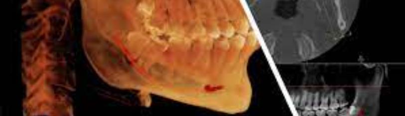
The ultrasonography of soft tissue structures of the mouth help in locating the soft tissue pathology involving those adipose tissue, muscles, tendons, or ligaments. It is an additional aid in diagnosing masseteric hypertrophy/hypotrophy, especially in patients suffering from oral submucous fibrosis. The new era of cone beam computed tomography (CBCT) has come up with a revolution in the field of dentistry. It assists dental professionals in the accurate location of pathology, impacted or embedded teeth, cysts, and tumors. Through CBCT, placement of implants has become quite easy as it helps in implant planning before the actual surgery. Also, surgical stents through 3-D printing have made the surgical placement of implants very precise and an easy task. Dental radiology with its various advancements has become a boon for the dental fraternity worldwide. Now, dental professionals can do more precise maxillofacial surgery as is aided by advanced dental radiographs.
Genesis Scientific Publication is licensed under CC BY-NC-ND 4.0![]()
![]()
![]()
![]()
