Role of Mesenchymal Stem Cells in Growth of Bone Grafting, Distraction of Bones and in Kummoona Chondro-Osseous Graft
Raja Kummoona*
Emeritus Professor of Craniomaxillofacial Surgery,Iraqi Board for Medical specializations,Baghdad, Iraq
*Corresponding author: Raja Kummoona, Emeritus Professor of Craniomaxillofacial Surgery,Iraqi Board for Medical specializations,Baghdad, Iraq
Citation: Kummoona R. (2024) Role of Mesenchymal Stem Cells in Growth of Bone Grafting, Distraction of Bones and in Kummoona Chondro-Osseous Graft. J Stem Cell Res. 5(2):1-04.
Received: August 02, 2024 | Published: August 10, 2024
Copyright© 2024 genesis pub Kummoona R CC BY-NC-ND 4.0 DEED. This is an open-access article distributedunder the terms of the Creative Commons Attribution-NonCommercial-No Derivatives 4.0 International License.,This allows others distribute, remix, tweak, and build upon the work, even commercially, as long as they credit the authors for the original creation.
Abstract
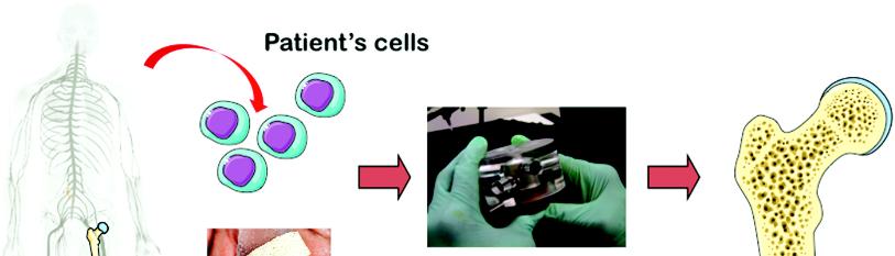
Mesenchymal stem cells play important role in reconstruction of temporomandibular joint (TMJ) by Kummoona chondro-osseous graft from iliac crest for restoration of normal biological function in growth, remodeling and repair.
Stem cells also play a role in growth of bone graft of lower jaw. These cells also participate in growth of distraction of jaws and short limb in children. These studies were supported by experimental studies on Rabbits to prove the ability of mesenchymal stem cells in growth of bones.
Histological studies showed enormous changes by formation of clot due to platelets aggregation, with releasing growth factor (PGF) and formation of healthy granulation tissues Mesenchymal stem cells were originated from bone marrow, released to periosteum and covering muscles.
There were no in histology of bone grafting and distraction technique except, distraction growth occurred by stretching of tissues and bone grafting by decortication of both graft and stump with rigiddifferences fixation
The role of mesenchymal stem cells in Kummoona chondro-osseous graft were used for replacement of damaged TMJ for restoration ofthe normal biological function of masticatory demands inof the TMJ., Ssurprising the cells of iliac bone converted from columnar type for growth in the body to multidirectional fashion in the TMJ because of environmental changes.
Keywords
Stem cells; Stem Cells; Distraction; Distraction; Graft.
Introduction
From our research and clinical studies for long time, we discovered that stem cells were playing important role in growth of Kummoona chondro-osseus graft, which been used for reconstruction of the temporomandibular joint (TMJ) in children.
Stem cells were also played an important role in growth of bone grafting and in distraction technique for elongation of jaws and short limbs in children. Stem cell a special cell able to create many different cell types like muscle and brain cells and able to have role to fix damaged tissues. There are three types of stem cells, blood stem cell, mesenchymal stem cells and neural stem cells.
During the last few years, stem cells have become therapeutic and known as stem cell therapy is playing important roles in regenerative medicine by promoting repair responses of diseased dysfunction. Stem cells might be able for repair of injured tissue, recently stem cells were applied for treatment of scoliosis of spine as new line of treatment instead of very complicated surgery of spine and consuming long time of surgery. This work made a revolution in treatment of spine scoliosis.
Kummoona Chondro-Osseous graft was advocated in 1986. The graft was harvested from iliac crest of growing child, aged between 4-6 years which consist of bone of 4-5cm with a cap of cartilage of 1cm width. This graft was used for reconstruction of damaged TMJ with sever deformity of the jaws and midface. These indicationschanges are were noticed in TMJ ankylosis and First Arch dysplasia Syndrome.
Application of chondro-osseus graft in reconstruction of the TMJ and the way of application of this graft made a revolution in science of reconstructive surgery of facial skeleton.
Reconstruction of the TMJ by biological element made a great challenge to all craniofacial surgeons for the last 7 decades for managements of ankylosed TMJ in children or for reconstruction of missing part of ramus and TMJ in cases of First Arch Syndrome which occurred due to early occlusion of embryonic stapedial artery which is the main nutrient vessel to First and Second branchial arches.
Methods of Reconstruction
Reconstruction by bone graft either immediately as urgent technique or later as elective procedure. Bone grafting required for reconstruction of jaws after radical resection of jaw tumors and wide resection of malignant tumors of orofacial region or in cases of post traumatic missile war injuries or for managements of limbs injuries. Our favorite site for bone grafting was the iliac crest because of high vascularity, shape and bulk. Bone graft harvested as cortico-cancellous type or bi cortical form, or as cancellous type used to promote healing of delayed type specially in long bones. Patients seek the best possible treatment with the most looking results and to restore the normal functional activity of the bone defect.
The ability to plane bone grafting comes only with long experience, skill and knowledge. In the early stage of practicing bone grafting the result can be hambling. Competence, skill and knowledge of bone grafting as a science can be achieved only through study every case for the patient needs and requirements based on general health of the patient.
Material, Methods and Results
Experimental studies were conducted by using on newly growing Rabbit as experimental model, the condyle excised and reconstructed by Kummoona Chondro-Osseus graft. Three months later the newly reconstructed joint was excised for histological studies. The specimens were immersed in 10% formalin with decalcified solution for 10 days. Sections slides were prepared after staining by(H&E) and studied by light microscope.
Four layers were observed, the first layer was an articulating layer containing dense and thick fibrocartilage tissues. The second layer contains several layers of round mesenchymal stem cells which represent the proliferative cell layer. The third layer consists of cells converted from columnar in the iliac crest to multidirectional fashion due to the functional demands of masticatory process with new environments simulating normal human condyle.
The fourth layer showing chondral ossification, chondrocyte was swollen and passes through series of changes of endochondral cells were converted to osteoid tissue, boney trabeculae with living osteoblast between bone marrow spaces were noticed.
The author published recently research study work by using Rabbits animal as experimental model for bone grafting and distraction [3,4]. The aim of these studies was to understand the cellular changes that occurred between the stump of jaw bone and in stretching bone in distraction.
Mesenchymal stem cells were playing important role in growth of bone graft and distraction of jaw. Growth factor (PGF) secreted released from platelets and mesenchymal stem cells were released from periosteum, bone marrow and covering muscles.
The histological and cytological changes in bone grafting and distraction was quite interesting and passes through series of changes. The cellular changes in both bone grafting and distraction of bone were more or less similar. It was initiated by formation of clot as first step fallowed by formation of healthy granulation tissues and large number of fibroblasts, tiny small vessels and osteoblasts were observed.
Experimental research proved to be of great importance to humans for better understanding of the cellular changes and the role of mesenchymal stem cells in bone grafting and distraction of jaws and long bones, not fully understood before [3,4].
From these studies and research, we observed the mechanism of cellular changes in bone grafting and distraction of bone technique by inducing mesenchymal stem cells are the same except distraction induced by stretching of tissue and bone was based on Illizarovf theory [5].
While successful bone grafting was achieved by maximum contact between bone graft and stump of bone segments with rigid fixation achieved by decortication of both bone graft and stump of the mandible by using plating or by soft stainless steel of 0.25mm with double 8 fixation was successful by our hands.
Conclusion
Mesenchymal stem cells played an important factor in growth of Kummoona Chondro-Osseous graft for reconstruction of the TMJ for restoring functional activity and growth of the condyle, ramus and midface. Also stem cells played an important factor in growth of bone grafting and distraction technique.
References
-
Kummoona R. (2020) The role of mesenchymal stem cells in Kummoona chondro-osseus graft for reconstruction of temporomandibular joint. J Stem Cell Res. 1(1):01
-
Kummoona R, Zayed A S. (2028) Reconstruction of lower jaw by iliac bone graft,experimental studies on Rabbits and role of mesenchymal stem cells. J Stem Cell Regen Biol. 4(1):20-24
-
Kummoona R, Jassim EAM. (2017) Distraction technique of lower jaw on Rabbits,experimental study and Research. J Stem Cell Regen Biol.3(2) : 158-62
-
Illizarof G A. (1988) The principle of Illizarof method. Bull Hosp J Dis Orthop Insit. 48(1):11-1.
-
McCarthy JG, Staffenberg DA, Wood RJ, Cutting CB, Grayson BH et al. (1995) Introduction of Intra-oral bone lengthening Device. Plast Reconstar surg. 4:978-81
-
Editorial Opinion. J Stem Cell Research.2024, editorial permission
Genesis Scientific Publication is licensed under CC BY-NC-ND 4.0![]()
![]()
![]()
![]()
