Retrorectal Hibernoma: a Rare Tumor with Unusual Presentation
Meriem Dehal1, Karim Haddad1, Salim Belkherchi2, Hadjer Ouahabi1, Fatiha Berrebiha1 and Salah Berkane1
1Bologhine hospital, Faculty of medicine of Algiers, Algéria
2Universitary and médical center of Bejaia, Faculty of medecine of Bajaia. Algeria
*Corresponding author: Berkane S, Head of visceral and oncologigal, Department of surgery, Bologhine hospital, Algeria
Citation: Dehal M, Haddad K, Belkherchi S, Ouahabi H, Berrebiha F, et al. (2023) Retrorectal Hibernoma : A Rare Tumor With Unusual Presentation. J Can Ther Res. 3(1):1-7.
Received: January 12, 2023 | Published: January 30, 2023
Copyright© 2023 genesis pub by Dehal M, et al. CC BY NC-ND 4.0 DEED. This is an open-access article distributed under the terms of the Creative Commons Attribution-NonCommercial-No Derivatives 4.0 International License., This allows others distribute, remix, tweak, and build upon the work, even commercially, as long as they credit the authors for the original creation.
DOI: https://doi.org/10.52793/JCTR.2023.3(1)-23
Abstract
Introduction
Hibernoma is a rare tumor that develops from fat cells. It is a benign tumor that grows slowly and rarely gives rise to particularly painful symptoms. The main differential diagnosis is liposarcoma. We report a case of a hibernoma with unusual location (retrorectal with gluteal development). Observation this is a 37-year-old woman with no significant history who presented with swelling of the left buttock that had been evolving for 5 years. The clinical examination revealed a swelling 20cm in large diameter that bulges at the level of the left buttock. This formation was painless, soft in consistency and the skin over it is of normal color and appearance. Digital rectal examination perceived a formation of soft consistency. This patient had no medical or surgical history. Soft in consistency and the skin over it is of normal color and appearance. Digital rectal examination perceived a formation of soft consistency. This patient had no medical or surgical history. A complete surgical resection was with simple post operative course. The histological study revealed a hibernoma. The patient is alive without recurrence at 8 months postoperatively.
Conclusion
This retrorectal localization of the hibernomaisvery rare. This tumor is currently well characterized with modern morphological examinations which allow a fairly precise diagnostic approach. Diagnosis may in some cases require cytopuncture.
Keywords
Hibernoma; Retrorectal location; Bisactumor
Introduction
Retrorectal tumors are a rare entity, often benign, asymptomatic, with a female predilection. They include a wide range of histological differentiation that can be benign or malignant [1]. Here, we report a case of retrorectal gluteal hibernoma, which is a rare benign tumor derived from brown fat, seen in fetuses and hibernating animals. It results in a mass of slow evolution that is not very painful. This type of tumor essentially poses a diagnostic problem with sarcomas.
Observation
This is a 37-year-old patient with no notable pathological history, who consulted for a painless formation of the left buttock. In the anamnesis, the patient specifies that this formation appeared for 5 years and gradually increased in size to reach a dimension of 20cm at present. It began to cause discomfort when the patient puts it in a sitting position. The patient was in good general condition and is a febrile. The clinical examination in the gynecological position and in lateral decubitus showed the following facts: a mass of soft consistency in the left buttock, 20 cm long, non-pulsatile, painless and not very mobile. The skin opposite had a normal appear ance both in its coloration and through its color and its palpation. There were no down stream neurovascular disorders or satellite adenopathies and hip mobility was normal. There was a depression of the inter-gluteal groove (Figure n° 1).

Figure n° 1: Clinical aspect of the tumour on examination.
The digital rectal examination found a normal looking anal margin as well as the tone of the anal sphincter. The finger perceived a formation of soft consistency in left laterorectal. The rectal wall was normal. The rest of the somatic examination was un remarkable. The biological examinations carried out were normal, in particular blood sugar levels, blood urea and creatinine, blood crasis and the inflammatory assessment.
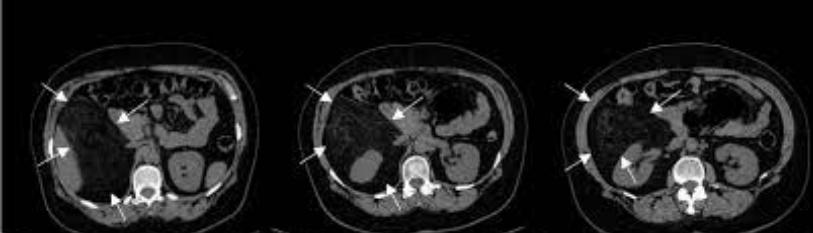
Pelvic ultrasonography (US) showed the existence of an inter-communicating cystic formation of the musculo-aponeurotic soft tissues of the left gluteal region and the posterior root of the under lying thigh. Abdomino pelvic computed tomography (CT Scan) revealed a septate, biloculated, thin-wall edcystic mass. She was enhanced with the injection of the contrast product. It was multi loculated with a component of the fatty plane of the left buttock of 170x70mm and a left perirectal component pushing back the rectum and the vagina of 100x70mm with integrity of the fat opposite (Figure n°2).

Figure n°2: Radiological aspect on CT Scan.
Magnetic resonance imaging (MRI) confirmed the presence of a multi-partitioned biloculated cystic mass delimiting cubicles and showed a bisac shape (Figure 3). It showed two cystic masses, one in the fatty plane of the buttock measuring 177x73 mm and the other extending towards the homo lateral is chio-rectal space of 200*70mm. This latter formation pushed the lower and middle rectum to the left and the vagina to the right without signs of infiltration. She had a persistent hyper signal on T1 on the FAT SAT sequences with lobular contours well limited by a clean wall and a hyper signal on T2, not enhance dafter contrast product (Figures n°3 and n°4).

Figure n°3: Radiological aspect on MRI (T1).

Figure n° 4: Radiological aspect on MRI (T2).
A spinal MRI ruled out the existence of an anterior sacral meaning ocele. Given these data, were trained the diagnosis of a benign lipomatous tumor and a surgical intervention with the aim of radical resection was scheduled. The installation of the patient was a gynecological position for a first perineal approach and a preparation for a possible abdominal approach if the first approach was not sufficient. A left perineal incision was made at the top of the formation with dissection flush with the tumor capsule was carried out step by step. This perineal incision was extended over the left buttock and towards the anococcygeal raphe. This approach continued slowly after separating the perineal muscles to reach the second part of the sutured formation in the left later orectal. This allowed us a complete excision of the formation. It should be noted that the latter was involuntarily perforated during the surgical maneuvers with issue of yellowish liquid. Ten milliliters of this liquid were sent for bacteriological study. This intervention ended with the placement of a Redon-type suction drain at the level of the tumor bed (left pararectal). Macroscopically, the tumor had a brownish appearance, trilobed on its internal face (Rectal face) and unilobed on its external face (Gluteal face) (Figure n°5 and n°6).

Figure n° 5: Tumor specimen after the resection (1).

Figure n° 6: Tumor specimen after the resection (2).
The bacteriological study of the fluid taken intraoperatively was negative. Microscopic examination showed a connective-muscular tissue seat of a benign mesenchymal proliferation arranged in nodules of variable sizes separated by fibrous septa; the tumor cells were arranged in a sheet of adipocyte cells with well-limited, eosinophilic, granular cytoplasm and sometimes optically empty appearance. Their nucleus is shifted to the periphery and regular. The stroma was reduced and fibrous. The over all appearance was in favor of a typical hibernoma. The postoperative follow-up was simple and the Redon drain was removed on the 4th post operative day. The patient was discharged on the 5th post operative day. At present, she is in good general condition with normal sitting position at 6 months post operative.
Discussion
Hibernomais a rare and unrecognized benign soft tissue tumor that develops at the expense of embryonic remains of brown fat. This tumor was first described in 1906 by Merkel [in 1]. These tumors have mainly been described for lesions occurring in the extremities and in the neck region, with only six cases of retroperitoneal hibernoma described in the literature [2-4]. To our knowledge, this is the second reported case of presacral hibernoma [5] and the first with gluteal development.
The term hibernoma was proposed in 1914 by Gery [in 1] because of the morphological similarity with the fat of hibernating mammals. It presents as a painless mass, with slow growth, sitting at various sites, the most frequent of which according to the largest series of studies by Furlong and his collaborators are: the thigh then the shoulder, followed by the back, the neck, thorax, upper limb and other sites [1]. Two hypotheses are put forward to explain the development of this tumour: the ectopic growth of brown fatty lobules and the migration fromb rown adipose tissue [6]. This tumor occurs between the ages of 30 and 40 with a slight female predominance. Slowly growing, it has a firm consistency and becomes painful in the event of compression of neighboring structures. Intense weight loss maya company the clinical picture. It is linked to excessive thermo genesis [7]. Indeed, unlike white fat, endowed with caloric reserve and thermal insulation capacities, brown fat is thermo genic through intense carbohydrate and lipid catabolism. After resection of the hibernoma, the weight returns to normal.
Modern morphological exploration allows a relatively easy diagnostic approach. On US, the hibernoma appears as a mass of homogeneous echo structure richly was cularized on Doppler [8]. On CT Scan, it is a heterogeneous hypodense lesion due to lipid, fibrous and vascular components [4]. Currently, MRI is the test of choice. It shows a more or less well-defined mass with a signal of intermediate intensity between that of striated muscle and subcutaneous fat. In T1, itis a signal of intensity discreetly lower than that of subcutaneous fat. In T2, the signal is most often heterogeneous. Upon injection of Gadolinium, contrast enhancement is intense, particularly in low-signal regions. Moreover, on fat saturation sequences, these tumors are not suppressed because of the nature of their lipids [9,10].
Liposarcoma is the number one differential diagnosis with hibernoma. This radiological aspect should lead to consider the hibernoma as a differential diagnosis of liposarcomas. A first biopsy is necessary if the tumor exceeds 5 cm on the MRI, especially in an elderly patient. The purpose of this biopsy is to eliminate a liposarcoma. However, it should be noted that percutaneous biopsy carries a hemorrhagic risk due to the hyper vascularization of the tumor [3]. Macroscopically, the hibernoma is a brownish tumor whose capsule is traversed by super ficial veins. Under microscopy, the typical appearance is made up of multi vacuolate cells with granular cytoplasm with small central nuclei.
Other differential diagnoses are: angiolipoma, spindle celllipoma, pleomorphic lipoma, lipoblastoma, elasto fibroma, clear cell sarcoma and alveolar sarcoma. Therapeutic management is only surgical with the type of complete resection without rupture of the fibrous capsule to avoid tumor recurrence. The pelvic location of the hibernoma in relation to the rectum, the surgical approach depends on the location, the size of the tumor and the surgical expertise.
Three main surgical approaches are used: anterior, posterior and combined. The anterior or trans abdominal approach is recommended for very large tumors, when there is suspicion of malignancy and when the lower limit of the tumor is above the third sacral vertebra. The posterior approach, first described by Paul Kraskein 1885, can be inter-sphincter, trans-sphincter para sacrococccygeal, trans-sacral, trans-sacrococcygeal, trans-anorectal, trans-vaginal. It is indicated in low tumors whose upper limit is less than S3, when there are signs of sacral involvement, in small tumors and when malignancy is not suspected.
A combined abdomino-sacral approach is recommended for larger lesions that extend both above and below S3, large masses, and if there is suspicion of malignancy [11]. In our case and given the benignity of the lesion and its development in the retro-gluteal region, a first perineal approach was decided with possible recourse to the combined approach in the abdomen if necessary. We were able to perform the tumor extirpation by pure perineal route. Therefore, the primordial question on the diagnostic level is the following: what is the nature of the tumour: is it a malignant tumor or a benign tumour? The sarcoma must be eliminated before the surgical therapeutic decision. For this purpose, an US or CT-guided biopsy may be essential in some cases. Apart from a preoperative biopsy, it is essential to respect the tumor without tumor opening because there is a risk of recurrence in this case. Due to the hyper vascularization of hibernomas, hemostasis must bemeticulous. The risk of degeneration is disputed. Only one case has been reported in the literature [12].
Conclusion
Hibernoma is a rare benign adipocyte tumor, often unrecognized and affecting young subjects with a slight female pre-dominance. It is necessary to know how to evoke it in front of a firm lipomatous tumor, richly was cularized, of brownish color which can be worrying during surgical excision. Presacral localization is very rare. Imaging (especially MRI) can rule out the differential diagnosis of liposarcoma. The curative treatment consists of a complete surgical excision which allows the histological confirmation of the diagnosis and avoids a recurrence.
References
- Furlong MA, Fanburg-Smith JC, Miettinen M. (2001) The morphologic spectrum of hibernoma: a clinicopathologic study of 170 cases. Am J Surg Pathol. 25(6):809-14.
- Sansom HE, Blunt DM, Moskovic EC. (1999) Large retroperitoneal hibernoma--CT findings with pathological correlation. Clin Radiol. 54(9):625-7.
- Cantisani V, Mortele KJ, Glickman JN, Ricci P, Passariello R, Ros PR, et al. (2003) Large retroperitoneal hibernoma in an adult male: CT imaging findings with pathologic correlation. Abdom Imaging. 28(5):721-4.
- Rigor VU, Goldstone SE, Jones J, Bernstein R, Gold MS, Weiner S. (1986) Hibernoma. A case report and discussion of a rare tumor. Cancer. 57(11):2207-11.
- Pandya A, Wasnik PA. (2011) Presacral hibernoma: Radiologic-pathologic correlation. Indian J Radiol Imaging. 21(4):270-3.
- Boudana D, Wolber A, Lesalle EM, Delaporte E, Martinot Duquennoy V. (2009) L’hibernome :une tumeur adipocytaire rare et méconnue. Ann Chir Plast Esthet. 56(2):156-9.
- Essadel A, Bensaid Alaoui S, Mssrouri R, Mohammadine E, Benamer S, Taghy A, et al. (2002) L’hibernome une cause rare d’amaigrissement massif. Ann Chir. 127(3):215-7.
- Seynaeve P, Montelmans L, Kockx M, Van Hoye M, Mathijs R. (1994) Case report 813 : hibernoma of the left thigh. Skeletal Radiol. 23(2):137-9.
- Dursuna M, Agayeva A, Bakira B, Ozgerb H, Eralpb L, Sirvancic M, et al. (2008) CT and MR characteristics of hibernoma: six cases. Clin Imag. 32(1):42-7.
- Ritchie DA, Aniq H, Davies AM. (2006) Hibernoma-correlation of histopathology and magnetic-resonance-imaging features in 10 cases. Skeletal Radiol. 35(8):579-89.
- Saxena D, Pandey A, Bugalia PR, Kumar M, Kadam R, et al. (2015) Management of presacral tumors: Our experience with posterior approach. 12:37-40.
- Alahyanea A, Bounaima A, Jahidb A, Janatia IM. (2006) Hibernome de l’avant-bras. Chir Mai. 25:166-8.
Genesis Scientific Publication is licensed under CC BY-NC-ND 4.0![]()
![]()
![]()
![]()
