Neurofibromatosis Type 1, Case Report of Surgical Treatment of Cervical C2 Kissing Neurofibromas
Marcel Sincari1, Francisco Cabrita2, Eduardo Mendes3 and Mark Sincari
1Centro Hospitalar Tondela-Viseu, Neurosurgery, PhD, Portugal
2CUF, Viseu, Neurosurgery , Portugal
3Centro Hospitalar, Ortopedy, Viseu, Portugal
4FMUC, Coimbra, Portugal
*Corresponding author: Marcel Sincari, Centro Hospitalar Tondela-Viseu, Neurosurgery, PhD, Portugal
Citation: Sincari M, Cabrita F, Mendes E and Sincari M. (2024) Neurofibromatosis Type 1, Case Report of Surgical Treatment of Cervical C2 Kissing Neurofibromas. J Neurol Sci Res. 4(2):1-9.
Received: August 12, 2024 | Published: August 27, 2024
Copyright©️ 2024 genesis pub by Sincari M, et al. CC BY-NC-ND 4.0 DEED. This is an open-access article distributed under the terms of the Creative Commons Attribution-Non-Commercial-No Derivatives 4.0 International License. This allows others distribute, remix, tweak, and build upon the work, even commercially, as long as they credit the authors for the original creation.
DOI: http://doi.org/10.52793/JNSRR.2024.4(2)-37
Abstract
Neurofibromatosis type 1 is an autosomal dominant genetic syndrome associated with numerous neoplastic and non-neoplastic manifestations affecting a variety of organ systems, including skin, eye, nervous system, skeleton, and endocrine and gastrointestinal tract. The diagnosis and clinical course of this disease is associated with a variety of different challenges. Postoperative management of surgically treated cases are even more difficult.
We present a case of a 18 years girl with NF1 with kissing NFBs of C2 root extreme cervical spine pressure and indications of myelopathy tended to precisely. The postoperative period was practically the follow up of complications and addressing them. The clinical result is very good, the patient’s satisfaction and life quality are also good and the overall follow up was 5 years.
Keywords
Neurofibroma; Myelopathy; Laminectomy.
Abbreviations
NF1: Neurofibromatosis type 1
NFBs: Neurofibromas
ACDF: Anterior cervical decompression and fusion
CSF: Cerebrospinal fluid
Introduction
The earliest descriptions of NF can be seen in Egypt papyrus of Ebers in 1500 before Christ, as well in artistic crops like Hellenistic statuette dated in 323 before Christ. More recent artistic descriptions are mosaic illustration of Ulise Aldrovandis, Monstrum Historia in 1592. The Quasimodo in the Victor Hugo´s novel Notre-Dame de Paris, published in 1832, is though that has NF [1].
One year later after Victor Hugo had published his Notre-Dame de Paris, in 1833, Friedrich Daniel von Recklinghausen (Virchow´s pupil) was born and had the first official interpretation of NF1 as an illness in 1882 on a report “On multiple cutaneous fibromas and their relationship with multiple neuromas” (Wilkins and Brody, 1971; Crump, 1981), he was the first to name the tumors neurofibromas [2].
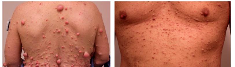
NF1 is estimated to occur in 1:3,000 live births. It is extremely variable in its clinical presentation from generalized expression of the disease (90% of the cases) to localized neurofibromas or café-au-lait spots [ 4]. According to another sources NF1 incidence is estimated of 1 in 2,700 and a prevalence of 1 in 4,500 [3].
NF1 is a monogenic desease with a pattern of autosomal dominant inheritance, with a high rate of de novo mutations of 42%. These mutations are believed to be responsible for the sporadic appearance of NF1 [5]. Penetrance is nearly complete before the age of 5 years, expressivity differs between affected individuals, even within the same consanguinity. There is no predilection for sex, race, or ethnicity [6,7]. NF1 is the most common tumor predisposition syndrome inherited in an autosomal dominant (100% penetrance) fashion [8]. The lifetime risk of malignancy in individuals with NF1 is estimated to be 59.6% [9], justifying the lifelong surveillance of patients with NF1 [10].
The diagnostic criteria for NF1 were defined in 1987 by the National Institute of Health Criteria Consensus Conference:
- Six or more spots of „cafe au lait” type more than 5 mm in diameter.
- Two or more neurofibromas or one plexiform neurofibroma.
- Freckles or skin discolorations in the places inaccessible for light (armpits, groins, pubic region).
- Two or more Lisch nodules on the iris.
- Characteristic skeletal changes.
- A first-degree relative suffering from NF1.
At least two criteria must be met for clinical diagnosis [11]. Specific skeletal changes of NF1 include congenital pseudarthrosis of the tibia, scoliosis, sphenoid wing dysplasia, rib penciling, and gracile bones [12]. Scoliosis can be considered the most common type of spinal deformity that it is present in 10% to 71% of cases [13]. According to another authors, spine deformities in NF1 including scoliosis, kyphosis and atlantoaxial instability are in 2- 36% of the patients [14].
Malignant transformation leads to the development of tumors as malignant peripheral nerve sheath tumor, or malignant triton tumor, sarcomas, like rhabdomyosarcoma, and other malignancies such as juvenile myelomonocytic leukemia or pheochromocytoma are also encountered in NF1 patients with increased frequency comparing to general population [15,16].
In 1992, the World Health Organization and National Neurofibromatosis Foundation declared that “although some clinicians have advocated routine screening scans for all patients with NF1, the utility of such screening has not been conclusively demonstrated” [17]. According to some sources, taking into account the rarity of complications that are usually symptomatic and easily detected during the clinical follow-up, screening investigations are not recommended [15,18]. Positron emission tomography/computed tomography is useful for the detection of malignant transformation of tumors in NF1 patients [19].
Case Report
34 years old lady, with NF1 diagnosed since the age of 18 years, admitted because of unstable gait. MRI revealed bilateral kissing C2 roots tumors with severe bilateral compression of the spinal cord, spinal cord is squeezed in between C2 bilateral tumors and C3 root left sided tumor (Figure 1). She was worked on, the extra and intradural developing cancers were taken out through C2, C3 laminectomy with ensuing dural remaking with nuchal tendon reap during the methodology. In the postoperative period she developed a huge, tense, subaponeurotic CSF collection (Figure 2), solved only after inserting the ventriculoperitoneal shunt with the use of navigation, because of small ventricles (Figure 3). Two years after the neurofibroma removal she referred neck pain and fixed position in anterior flexion of the head. X-Ray, MRI diagnosed post laminectomy regional kyphosis C2-C3 with anterior luxation. She was selected for circumferential arthrodesis (ACDF C3-C4, C4-C5, posterior mass lateral fixation C2- C6 on the right side, C2, C3, C5, C6 on the left side) with good recovery and pain relief (Figure 4). Three years after she started to be bothered by upper limb paresthesia and MRI revealed initial cervical syringomyelia. One year later her gait was progressively more unstable and MRI showed significant syringomyelia progression (Figure 5) and next surgery was performed: siringopleural shunt insertion trough upper thoracic laminectomy Th1, Th2 and unilateral intralaminar fixation C7-Th3 was performed at the end of the surgery (Figure 6). The choice to fix C7-Th3 with intralaminar one-sided screws was directed by past involvement in cervical shakiness and feeble muscles of the patient. The cranial hand of the proximal T shaped shunt is placed inside the syrinx cavity and the caudal part was positioned in the subdural space (Figure 7), the distal part was inserted in the pleural space through the intercostal space. The postoperative period was uneventful, the gait improved significantly. The follow-was 5 years, she is doing well with very few unspecific complaints.

Figure 1: MRI revealing bilateral C2 root tumor with severe bilateral spine cord compression and C3 unilateral tumor.


Figure 2: MRI and CT scan showing huge, postoperative pseudo meningocele, no signs of hydrocephalus, no signs of residual tumors.

Figure 3: Serial CT scan after ventriculoperitoneal shunting, meningocele diminished on serial CT scan.

Figure 4: MRI revealing upper cervical post laminectomy regional kyphosis, syringomyelia, no signs of meningocele. X-Ray before and after 360-degree arthrodesis.

Figure 5: Serial MRI with 1-year time difference, showing syringomyelia progression to upper thoracic spine cord.

Figure 6: CT scan, X-Ray after syringopleural shunting.

Figure 7: Intraoperative picture, the cranial hand of the proximal part of T shaped shunt is placed inside the syrinx cavity and the caudal part is positioned in the subdural space.

Figure 8: MRI 1 year after siringo-pleural shunting, reveal no intramedullary cavity.
Discussion
The most common tumor found in NF1 patients are NFBs, followed by plexiform NFBs, malignant peripheric nerve sheath tumors, and glial tumors [20]. Contrary to NF2, the tumors are less frequently found in the intradural spinal compartment in NF1 (less than10% of the cases), with most of them located laterally to the neuroforamina [21].
Like in the case described here, NFBs are localized bilaterally in two nerve roots at the same spinal level, resulting in significant cord compression. These lesions called “kissing neurofibromas”, in most cases associated with progressive myelopathy in the cervical spine, usually with surgical indication to relieve cord compression. In the literature is mentioned that decompressive procedures in the cervical spine in NF1 patients will ultimately cause some degree of deformity, even in adult patients [22], like what happened in our case.
The best treatment of malignant peripheric nerve tumors is complete excision. Adjuvant radiation and chemotherapy is reported with unclear success rates. Neurofibromatosis-1 patients with MPNSTs have a 5-year survival rate of approximately 21% [23]. Hydrocephalus may be an unexpected postsurgical sequela in these patients [24]. The subaponeurotic CSF collection after removal of C2 tumors in our opinion is like external hydrocephalus, that regressed only after ventriculoperitoneal shunting.
Conclusion
Despite our wide knowledge concerning NF1 there are still numerous clinical and surgical challenges to be solved. Surgical removal is the principal treatment for neurofibromas, with a high recurrence rate after partial removal of large plexiform neurofibromas. In the case of NF1-related tumors, there is no consensus with regard to the treatment strategy due to the multiple pathways involved in the growth of NF1 tumors [25,26].
More relevant spinal complications of NF1 include pseudarthrosis, bleeding, hematoma formation. Progression of the deformity may also be observed, as well as, dural leaks [27]. Based on a group of 22 patients treated surgically, in retrospective analysis, it was concluded that early stabilization of the cervical spine prevents late deformity of the patients with NF1 [22]. Our patient suffered a series of complications, more relevant being CSF flow disturbances, huge tense subaponeurotic collection solved with shunting and progressive syringomyelia, in spite of good functioning of ventriculoperitoneal shunt, that was resolved with siringo-pleural shunting. Another major complication was cervical post-laminectomy kyphosis, that could be avoided if initial fixation would be done at the first surgery. We would like to stress that preoperative planning and tight follow up of the surgically treated patients withnNF1 is an important issue, that permits to asses and address properly the evolutive complication and improve the patient’s life quality.
Funding
None
Conflict of Interest
None
References
Genesis Scientific Publication is licensed under CC BY-NC-ND 4.0![]()
![]()
![]()
![]()
