Nasal Mucosal Leishmaniasis: a Case Report
Yasmine Rkiek* Ouiame El Jouari and Salim Gallouj
Department of Dermatology, University Hospital Center of Tangier, Tetouan Al Hoceima, Faculty of Medicine and Pharmacy Tangier Abdelmalek Essaadi University, Tangier, Morocco
*Corresponding author: Yasmine Rkiek , Department of Dermatology, University Hospital Center Tangier, Tetouan Al Hoceima, Faculty of Medicine and Pharmacy Tangier Abdelmalek Essaadi University, Tangier, Morocco
Citation: Rkiek Y, Jouari OE, Salim Gallouj. Nasal Mucosal Leishmaniasis: A Case Report. J Clin Pract Med Case Rep. 2(1):1-5.
Received: January 30, 2025 | Published: February 10, 2025.
Copyright© 2025 genesis pub by Rkiek Y, et al. CC BY-NC-ND 4.0 DEED. This is an open-access article distributed under the terms of the Creative Commons Attribution-Non Commercial-No Derivatives 4.0 International License. This allows others distribute, remix, tweak, and build upon the work, even commercially, as long as they credit the authors for the original creation.
DOI: https://doi.org/10.52793/JCPMCR.2025.2(1)-22
Abstract
Leishmaniasis is a parasitic disease with various forms, including visceral, cutaneous, and mucocutaneous types In Morocco, cutaneous leishmaniasis is endemic, while mucocutaneous leishmaniasis (MCL), particularly nasal mucosal involvement, is rare. We report a case of nasal mucosal leishmaniasis in a 55-year-old immunocompetent woman with a 2-year history of a treatment-resistant papular lesion. Diagnosis was confirmed after biopsy, revealing Leishman bodies, and the patient was successfully treated with intramuscular meglumine antimoniate, showing significant improvement within the first week. Nasal mucosal leishmaniasis is exceptionally uncommon and responds well to standard treatment in immunocompetent patients. Early diagnosis and appropriate treatment are crucial for a favorable outcome, even in atypical cases like this one.
Keywords
Leishmaniasis; Nasal Mucosal; Parasitic disease
Introduction
Leishmaniasis is a parasitic disease with a wide range of clinical manifestations, including visceral leishmaniasis (VL), cutaneous leishmaniasis (CL), and mucocutaneous leishmaniasis (MCL). In Morocco, the visceral and cutaneous forms represent significant public health concerns in certain regions, while the mucocutaneous form remains rare, with only a few cases reported in the literature [1]. Here, we present a case of nasal mucosal leishmaniasis in an immunocompetent woman. The main objective of this study is to investigate the potential of using iPSCs to generate HIV-resistant immune cells for autologous transplantation to reconstitute the immune systems of HIV-positive individuals.
Observation
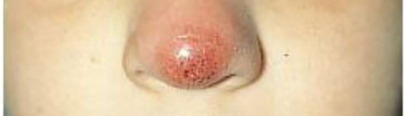
A 55-year-old female patient, with a history of pulmonary tuberculosis treated 4 years ago, presented with a treatment-resistant papular lesion on the nasal mucosa that had been evolving for 2 years. Clinical examination revealed a papule approximately 1 cm in diameter in the nasal vestibule. Dermoscopy showed yellowish and whitish scales on an erythematous base, with a characteristic teardrop appearance and spotty vascularization. Examination of other systems was unremarkable, with no evidence of fever or signs of systemic involvement.
Initial parasitological examination was negative, and a first biopsy of the nasal vestibule revealed a chronic inflammatory infiltrate. A second biopsy confirmed the diagnosis of leishmaniasis of the nasal vestibule, with the presence of Leishman bodies mixed with histiocytes and neutrophilic polymorphonuclear cells.
The patient was treated with intramuscular injections of meglumine antimony, showing significant improvement within the first week.
Discussion
The particularity of this case lies not only in the rare localization of leishmaniasis in the nasal mucosa but also in the favorable outcome observed following treatment. Mucocutaneous leishmaniasis (MCL) is generally uncommon, and its occurrence in the nasal mucosa is exceptionally rare [1]. In Morocco, localized cutaneous leishmaniasis (CL) remains endemic in several regions, whereas MCL is more prevalent in South and Central America [1]. The exact mechanism for the localized involvement of the nasal mucosa remains unclear, but it could be explained by the physical characteristics of the nasal region [2].
Leishmaniasis is transmitted by sandflies (genus Phlebotomus), which bite and introduce the Leishmania parasite into the host [3]. While sandflies typically bite exposed areas of the skin, the nasal mucosa, particularly the vestibule, is less likely to be affected [2]. This is likely due to the airflow during inhalation and exhalation, which could deter the sandflies from biting the area. The nasal mucosa, with its unique anatomical and physiological features, may create an environment less conducive to parasite establishment compared to other mucosal surfaces, such as those in the mouth or pharynx. This anatomical phenomenon may help explain the rarity of mucosal leishmaniasis in this location.
Clinical diagnosis of mucosal leishmaniasis, especially when it presents in isolation, can be quite challenging [4]. Symptoms such as a persistent, treatment-resistant papule in the nasal vestibule often mimic other dermatologic or infectious conditions, making it difficult to suspect leishmaniasis without further diagnostic evaluation. In this case, the use of dermoscopy was crucial, revealing characteristic features of Leishmania infection, such as yellowish and whitish scales on an erythematous base with teardrop-shaped lesions and spotty vascularization. While dermoscopy is not commonly used for diagnosing leishmaniasis, this technique can offer important clues, particularly when dealing with atypical presentations [5]. Further histopathological examination and biopsy, which are essential for definitive diagnosis, revealed the presence of Leishman bodies, confirming the diagnosis of leishmaniasis (Figure 1,2).

Figure 1: Nasal leishmaniosis lesion.

Figure 2: Improvement after treatment.
Unlike the mucocutaneous leishmaniasis seen in the Americas, which is typically more destructive and associated with significant morbidity due to its invasive and progressive nature, nasal mucosal leishmaniasis in Morocco does not usually result in the same degree of tissue destruction. The lesion in our patient was confined to the mucosal surface, without significant extension into the deeper nasal structures. This could be attributed to differences in the Leishmania species or strains involved. In Morocco, the causative agent is most often Leishmania major or Leishmania tropica, both of which tend to cause localized and self-limiting infections [6]. Conversely, in the New World, Leishmania braziliensis, which causes mucocutaneous leishmaniasis, is more aggressive, leading to widespread mucosal and skin involvement with potential for disfigurement. In our case, the good response to treatment with meglumine antimoniate, a first-line therapy for leishmaniasis, further supports the less virulent nature of the parasite in this region.
Meglumin antimoniate remains the gold standard treatment for leishmaniasis [7]. and its use in this case led to rapid improvement, corroborating the high efficacy of this drug in the treatment of leishmaniasis of the mucous membranes. The typical treatment regimen for mucocutaneous leishmaniasis involves a course of systemic antimonial therapy, with dosage and duration depending on the clinical severity and localization of the lesions. While the side effects of antimony-based treatments can be significant, including hepatotoxicity and nephrotoxicity, these drugs continue to be the mainstay of therapy due to their effectiveness [8, 9]. Alternatives such as miltefosine and amphotericin B are also available but are generally reserved for more severe cases or when antimony compounds are not tolerated [10].
A noteworthy point in our case is the patient's immunocompetence, which likely contributed to the favorable outcome. Leishmaniasis tends to affect immunocompromised individuals more severely, leading to disseminated or chronic infections that are difficult to treat. Immunocompromised patients, particularly those with HIV/AIDS or those undergoing immunosuppressive therapy, may exhibit more extensive and recalcitrant mucocutaneous involvement. In contrast, immunocompetent individuals like our patient typically exhibit a more localized and self-limited form of the disease.
The outcome of this case also underscores the importance of early diagnosis and prompt treatment. Early detection of leishmaniasis can significantly reduce the risk of complications, including the destruction of mucosal tissues, which can result in permanent disfigurement, particularly when nasal or oral mucosa are involved. In regions where leishmaniasis is endemic, clinicians should be vigilant in considering the diagnosis in patients presenting with persistent, atypical lesions, especially when they are resistant to standard treatments for other conditions.
Conclusion
Although nasal mucosal leishmaniasis is extremely rare, this case highlights the possibility of its occurrence in immunocompetent patients and the efficacy of standard treatment with meglumine antimoniate. Early diagnosis and appropriate management can lead to favorable outcomes, even in rare and atypical forms of the disease.
Acknowledgements
None
References
Genesis Scientific Publication is licensed under CC BY-NC-ND 4.0![]()
![]()
![]()
![]()
