Mandibular Retiform Hemangioendothelioma: Exploration of a Rare Localization
Anouar Titou*, Taha Jazzar, Hind Boukaaba, Mohammed Essioti and Dounia Kamal
Department of Reconstructive and Maxillofacial Surgery, Hassan II University Hospital of Fez, Sidi Mohamed Ben Abdellah University, Faculty of Medicine, Pharmacy, and Dentistry, Fez, Morocco.
*Corresponding author: Anouar Titou, Department of Reconstructive and Maxillofacial Surgery, Hassan II University Hospital of Fez, Sidi Mohamed Ben Abdellah University, Faculty of Medicine, Pharmacy, and Dentistry, Fez, Morocco.
Citation: Titou A, Jazzar T, Boukaaba H, Essioti M, Kamal D. Mandibular Retiform Hemangioendothelioma: Exploration of a Rare Localization (A Case Report). Genesis J Surg Med. 1(1):1-5.
Received: January 1, 2022 | Published: January 15, 2022
Copyright© 2022 genesis pub by Titou A et al. CC BY- NC-ND 4.0 DEED. This is an open-access article distributed under the terms of the Creative Commons Attribution-Non-Commercial-No Derivatives 4.0 International License. This allows others distribute, remix, tweak, and build upon the work, even commercially, as long as they credit the authors for the original creation.
DOI: https://doi.org/10.52793/GJSM.2022.1(1)-1
Abstract
Introduction: Retiform hemangioendothelioma (RH) is a rare vascular tumor characterized by its distinctive vascular architecture and indolent clinical course. While it primarily occurs in the limbs, its presence in the mandibular symphysis is exceptional.
Case Report: We present the case of a 60-year-old woman with a painful, firm lesion located in the mandibular symphysis. Radiographic examinations revealed radiolucent areas with cortical destruction. A cervicofacial CT scan showed the extent of the lesion, including a submental lymph node.
Surgical management involved wide excision and bilateral neck dissection, followed by reconstruction using a pectoralis major myocutaneous flap and a rib graft. Histopathological examination confirmed the diagnosis of RH.
Discussion: RH is a rare tumor that should be considered in the differential diagnosis of atypical vascular lesions in the maxillofacial region. Management typically involves wide excision and functional reconstruction, with long-term follow-up required due to the risk of local recurrence.
Conclusion: This case illustrates the rarity of RH in the mandibular region and highlights the importance of a multidisciplinary approach, including complete resection and appropriate reconstruction. Long-term monitoring is essential to prevent recurrence.
Keywords
Retiform hemangioendothelioma; Mandibular Symphysis; Vascular Tumor; Neck Dissection; Pectoralis Major flap; reconstruction
Introduction
Retiform hemangioendothelioma (RH) is a rare vascular tumor first described by Calonje et al. in 1994 [1]. It is classified as an intermediate-grade neoplasm, falling between benign hemangiomas and malignant angiosarcomas, with a known potential for local recurrence but a low risk of metastasis [2]. RH predominantly affects the extremities of young adults and has only rarely been reported in the head and neck region [3]. Histopathologically, RH is characterized by elongated, arborizing blood vessels arranged in a pattern resembling the rete testis [4]. In this report, we describe a unique case of RH involving the mandibular symphysis, an extremely rare site, in a 60- year-old woman presenting with a painful and indurated lesion.
Case Report
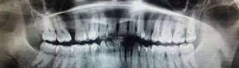
A 60-year-old woman presented with a two-month history of intense pain and swelling in the chin area. She had no significant medical history and denied any prior trauma. Upon examination, a firm, indurated mass was noted in the mandibular symphysis, tender upon palpation. The overlying mucosa appeared intact without ulceration, but a small submental lymph node was palpable (Figure 1). Panoramic radiographs revealed multiple radiolucent areas in the symphyseal region (Figure 2), with evidence of cortical bone destruction. A cervicofacial CT scan confirmed a heterogeneous mass within the symphysis (Figure 3), with bone erosion extending into the adjacent soft tissues. Enlarged submental lymph nodes were suggestive of reactive adenopathy.
Surgical management involved a wide local excision of the tumor with bilateral neck dissection. Reconstruction of the mandibular defect was performed using a pectoralis major myocutaneous flap and a rib graft. The patient recovered well postoperatively and was discharged two weeks later.
Histopathological analysis confirmed the diagnosis of RH. Microscopy revealed an irregular network of elongated and branching vessels lined by hobnail endothelial cells. Immunohistochemical staining was positive for CD31 and CD34, further supporting the diagnosis [5, 6], (Figure 4). At the six-month follow- up, the patient showed no signs of recurrence, with a well-healed surgical site and no further lymph node enlargement. Regular follow-up visits are scheduled to monitor for potential recurrence.
Discussion
RH is an exceedingly rare vascular neoplasm, with fewer than 50 cases reported in the literature [7]. While RH is most commonly observed in the limbs, its occurrence in the mandibular symphysis is exceptional. The presentation of a painful and indurated mass with cortical bone destruction is unusual for typical vascular tumors, making this case noteworthy. The imaging studies played a crucial role in assessing the extent of the tumor, revealing its involvement with adjacent soft tissues and highlighting the necessity for a comprehensive surgical approach [8].
Wide local excision was essential for achieving local control of the tumor, a strategy supported by the literature on RH management. Neck dissection, although more commonly associated with malignant tumors, was performed due to the suspicious submental lymph nodes. However, subsequent histopathology confirmed their reactive nature, and no metastatic spread was identified [9].
Reconstruction of the mandibular defect using a pectoralis major myocutaneous flap combined with a rib graft offered both functional and aesthetic benefits, a choice corroborated by numerous studies highlighting the reliability of this flap in complex head and neck reconstructions [10]. This approach allowed for optimal coverage of the defect and restoration of mandibular contour.
Histologically, RH is distinct from other vascular tumors, particularly angiosarcoma, due to its characteristic retiform (net-like) pattern of arborizing vessels and hobnail endothelial cells [11]. The immunohistochemical profile, including positivity for endothelial markers CD31 and CD34, further aids in differentiating RH from more aggressive malignancies [12].
Although RH is classified as a tumor with low metastatic potential, local recurrence is common, with studies reporting rates as high as 50% [13]. Therefore, long-term follow-up is critical, even in cases where the initial treatment appears successful. In our patient, there has been no recurrence to date, but vigilance remains paramount due to the high recurrence risk associated with this tumor [14].
Conclusion
Retiform hemangioendothelioma is an extremely rare vascular tumor that presents unique diagnostic and therapeutic challenges, particularly when located in the mandibular symphysis. This case highlights the importance of a multidisciplinary approach involving wide excision, neck dissection, and functional reconstruction with a pectoralis major myocutaneous flap and rib graft. The successful management of this case adds valuable insight into the limited body of literature on RH in atypical locations, underscoring the need for continued surveillance to detect and manage recurrences early [15].
Conflict of Interest
The authors declare no conflicts of interest.
Authors' Contributions
All authors participated in the patient's care and in the writing of the article. All authors have read and approved the final version of the manuscript.

Figure1: Clinical images (exobuccal and endobuccal) showing a normal appearance of the skin andoral mucosa in relation to the symphyseal lesion.

Figure 2: Panoramic radiographs revealed multiple radiolucent areas in the symphyseal region.

Figure 3: CT scan confirmed a heterogeneous mass within the symphysis, with bone erosionextending into the adjacent soft tissues.

Figure 4: Retiform hemangioendothelioma composed of long arborizing blood vessels with a hobnailappearance of the lining cells.
References
Genesis Scientific Publication is licensed under CC BY-NC-ND 4.0![]()
![]()
![]()
![]()
