Incidental Finding of Migrated Intra-Uterine Device during Elective Robotic-Assist Laparoscopic Hysterectomy
Cash C Sterling, Francis Lake and James Chambers*
Department of Surgery, Northeast Georgia Heath System, Braselton, GA, USA
*Corresponding author: James Chambers, Department of Surgery, Northeast Georgia Heath System, Braselton, GA, USA
Citation: Sterling CC, Lake F, Chambers J. (2022) Incidental Finding of Migrated Intra-Uterine Device during Elective Robotic-Assist Laparoscopic Hysterectomy. 3(1):1-4.
Received: January 07, 2022 | Published: January 31, 2022
Copyright© 2022 genesis pub by Sterling CC, et al. CC BY-NC-ND 4.0 DEED. This is an open-access article distributedunder the terms of the Creative Commons Attribution-NonCommercial-No Derivatives 4.0 International License.,This allows others distribute, remix, tweak, and build upon the work, even commercially, as long as they credit the authors for the original creation.
DOI: https://oi.org/10.52793/ACMR.2022.3(1)-24
Abstract
Migration of Intra-uterine devices (IUDs) a rare, yet serious complication associated with a well-accepted means of contraception. IUDs have long been a safe and effective option for women of child-bearing age to prevent pregnancy. We present a 48-year-old female with known symptomatic uterine fibroids undergoing a robot-assist laparoscopic hysterectomy in the elective setting. She was found to have a previously placed IUD that migrated through her uterus and was adherent and invading into her rectum. This IUD was removed without complication and without perforation of bowel. Surgical retrieval of IUD perforation should always be considered whether symptomatic or not, to prevent serious complication. Laparoscopic removal is the option of choice, but laparotomy may be required.
Introduction
Intra-uterine devices have been considered a first-line option for reversible contraception according to the World Health Organization (WHO). Between 2015 and 2017, over 14% of women aged 15 to 44 who used contraception chose IUDs and those numbers continue to rise. IUDs work by preventing the ovum and sperm from fertilization and are greater than 99% effective at preventing pregnancy. Migration of IUDs through the uterine wall is an infrequent complication with a reported incidence between 1.3 and 1.6 per 1000 placed. This report describes the incidental finding, removal, and management of a migrated IUD causing serosal injury to the rectum during elective robot-assist laparoscopic hysterectomy [1-4].
Case Report
A 48-year-old Caucasian female with history of indicative uterine fibroids introduced for elective absolute stomach hysterectomy with reciprocal salpingectomy. She is G2P0-0-2-0 (AB x2) with worsening menorrhagia and dysmenorrhea described as extremely heavy and painful. She has no history of uterine masses or family history of gynecologic malignancy [5-8]. Prior pelvic ultrasonography was significant for uterine leiomyoma, but no IUD was detected. An IUD had been placed eleven years prior and was thought to be lost through vaginal migration (Figure 1,2).

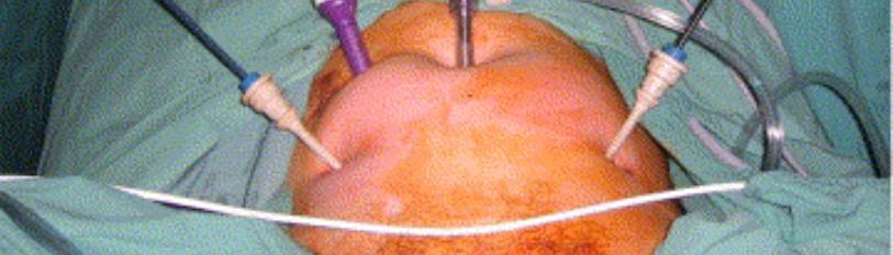
Figure 1: Intra-op photos of IUD found intraperitoneal.

Figure 2: Intra-op photo of colonoscopy showing no luminal defect.
During standard dissection of the left broad ligament of the uterus using a da Vinci Xii robot, strings of an IUD were identified. The IUD was found fully expulsed through the uterus in the cul-de-sac and embedded into the serosa of the adjacent rectum. Extensive endometriosis was noted within the pelvis including involvement of bilateral ovaries and in the cul-de-sac. Robot-assist laparoscopic hysterectomy was completed with excision of endometriosis and bilateral Salpingo-oophorectomy. Intraoperative consultation to General Surgery was performed. The patient was set in the left sidelong decubitus position, the butt-centric area was analyzed, a rectal test was played out, the sigmoidoscope was embedded and progressed without trouble to a distance of 25 cm. The IUD was removed laparoscopically. A leak test was performed to assess for perforation which was not seen. The area of concern was reviewed intently, and there was a little area of disintegration or tension injury to the serosa which was fixed with a 3.0 vicryl stitch in a basic intruded on manner. Another leak test was performed, and no leak was observed. No evidence of rectal perforation was observed. The procedure was completed; the patient was intubated and was taken to the post-operative area in stable condition.
The patient was admitted to the hospital for close observation and intravenous antibiotics. Diet was advanced on post-operative day one. The patient remained afebrile without concerning findings of bowel perforation including normal white blood cell count, benign abdominal exam, and normal passage of flatus and bowel movement. She was discharged on post-operative day two with a regular diet and a 5-day course of oral ciprofloxacin and metronidazole. She was seen in clinic on post-operative day twenty-six and was fully recovered. Colonoscopy was planned for 6-8 weeks.
Discussion
Intra-uterine device use remains a safe, effective, and popular method of reversible contraception. Intra-uterine device migration is a rare, but potentially serious complication associated with this reversible contraception method. Incidentally found foreign bodies should be removed urgently before bowel, urinary bladder, or other organ penetration occurs. Although this patient did not experience an adverse outcome associated with this rare complication, morbidity does occur. We conclude that safe removal of migrated IUDs found incidentally during laparoscopic surgery can be done and intra-operative endoscopy should be performed to assess the rectum and colon in similar cases.
Declaration of Patient Consent
Patient grants consent for publication.
References
- Li R, Li H, Zhang J, Li H. (2021) Rectum migration of an intrauterine device. J Minim Access Surg. 17(1):113.
- Ye H, Huang S, Zhou Q, Yu J, Xi C, et al. (2018) Migration of a foreign body to the rectum: A case report and literature review. Medicine. 97(28):e11512.
- Ma GW, Yuen A, Vlachou PA, de Montbrun S. (2016) An unconventional therapeutic approach to a migratory IUD causing perforation of the rectum. J Surg Case Rep. 2016(2):rjw004.
- DiPaola L, Wonaga A, Dardanelli M, Viola L. (2017) Intrauterine device in the rectal cavity. Rev Esp Enferm Dig. 109(4):290.
- Han X, Yang H. (2021) Successful endoscopic management of 3 cases of translocated intrauterine devices: a case report. Ann Palliat Med. 10(2):2371-78.
- Medina TM, Hill DA, DeJesus S, Hoover F. (2005) IUD removal with colonoscopy: a case report. J Reprod Med. 50(7):547-49.
- Shute L, Pidutti J, Trepman E, Burnett M, Embil JM. (2020) Rectal Perforation by an Intrauterine Device Leading to Fatal Intra-Abdominal Sepsis and Necrotizing Fasciitis. J Obstet Gynaecol Can. 43(6):760-2.
- Al Sahaf MA, Bseiso BF, Al-Momen SA, Meshikhes AN. (2019) Endoscopic removal of an incidentally discovered intrauterine contraceptive device eroding into the rectum. BMJ Case Rep. 12(9):e231410.
Genesis Scientific Publication is licensed under CC BY-NC-ND 4.0![]()
![]()
![]()
![]()
