Development of Hassall’s Corpus Cells
Gursharanjeet Singh Mann1* and Kunal Joon2
University, NIIMS greater Noida
*Corresponding author: Gursharanjeet Singh Mann. University, NIIMS greater Noida
Citation: Mann GS, and Joon K. Development of Hassall’s Corpus Cells. Genesis J Microbiol Immunol.1(1)-6.
Received: April 02, 2025 | Published: April 20, 2025
Copyright©2025 by Mann GS, et al. All rights reserved. This is an open access article distributed under the terms of the Creative Commons Attribution License, which permits unrestricted use, distribution, and reproduction in any medium, provided the original author and source are credited.
Abstract
Development of Hassall’s corpus cells is independent of the Age and these are required for T cell development and giving. The property of the self-antigen tolerance and the most common type of hassle corpus cells is a mosaic type found during early stage of development of hassle corpus cells.
Keywords
Hassall’s corpus cells; Mosaic type; Calcified Hassall’s corpus cells; Fifth brachial arch.
Introduction
Hassall’s corpus cells developed from type 6 thymic epidermal cells and these develops the receptor for the T cells and give the ability of the self-antigen recognition and preventing the autoimmune diseases [1].
Experiment 1
Aim: to Observed Development of Hassall’s corpus cells
Methodology: slides for the Development of thymus
Observation: Thymus was observed at different Stages of Development [2].

Figure 1: Photomicrograph of foetus thymus at 9th week of gestation showing capsule, lobulation, Development of cortex and medulla.

Figure 2: slide of the foetal thymus at 12 weeks showing lobulation, capsule, cortex, medulla and trabeculae containing blood vessels.

Figure 3: slide of foetal thymus at 14 week of gestation showing mosaic pattern of Hassall’s corpus cells.

Figure 4: Oil immersion photomicrograph of foetal with leishman’s stain showing lymphocytes, macrophage.

Figure 5: Photomicrograph of foetal thymus at 17 weeks of gestatn.

Figure 6: photomicrograph of foetus thymus at 18 weeks of gestation showing Hassall’s corpus cells Development.

Figure 7: At 24 weeks of gestation foetus thymus showing Hassall’s corpus cells at the medulla of thymus.

Figure 8: Showing further Development of Hassall’s corpus cells.

Figure 9: full Development of Hassall’s corpus cells.
Result
- Development of Hassall’s corpus cells start at 14 weeks of gestation and the further formation of Hassall’s corpus cells occur age independent manner.
- The greatest Development progression and main cells tissue organization of Hassall’s corpuscle was observed between 6 and 10 lunar months.
Experiment 2
Aim: to Observed ectopic thymus tissue from different individual died in different.
Cases
Material: slides of the ectopic thymus tissue (of 38 years mother, 14 years old boy and 10 years old boy) [3].
Observation

Figure 1: It’s a picture of normal position or ectopic thymus. showing reactivity of Hassall’s corpuscles Showing same immune reaction as in normal thymus.

Figure 2: Complete involution of thymus observed in boy died due to hunger. Small number of lymphocytes and involution of thymus [4].

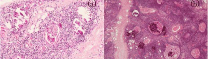
Figure 3: Mosaic pattern of Hassall’s corpuscles is observed in couples of mother/boy (a,b). And mother and daughter (c,d). This pattern is seen in new-born [5].

Figure 4: The Hassall’s corpuscles with calcification and thymic involution. A) Many numbers of Hassall’s corpuscles from 4 years old girl who died due to sepsis are change into calcification, and involution of thymus B) partial calcification of Hassall’s corpuscle were observed in a 14 years old boy died due to subdural bleeding after two weeks of head injury [6].
Result
Types of Hassall’s corpuscle
Immunopositive Hassall’s corpuscle
Immunopositive Hassall’s corpuscle: these basically select the receptor for T cells and regulate them and generate the thymosin to generate more T lymphocytes and decrease with the increase in the age Mechanism of generation of thymosin in Immunopositive Hassall’s.
Corpuscle
Step 1: Dissolving of the thymic epidermal cells or lymphocytes by macrophage.
Step 2: Taking of protein and breaking it down into peptide by type 6 epidermal reticular cells and conversion it into thymopoeitin and thymosin.
Step 3: Realising of thymopoeitin and thymosin by the mast cells.
Immmunonegative Hassall’s corpuscle: these basically produce immunosuppressive agent for cleaning of clone T cells.
Steps of production of immunosuppressive secretions
Step 1: first lymphocytes react with lymphocytes components present in Hassall’s corpuscle
Step 2: starts a immune reaction and leads to stimulation of type 6 epidermal reticular cells leads to generation of peptide.
Step 3: conversion of peptide into cortisol and leads mast cell to production of cortisol and elimination of cloned cells.
Discussion
In this we discussed about the Development of Hassall’s corpuscle and how they developed in stress situation and their development from the infancy and then we discussed about the types of Hassall’s corpuscle and their mechanism of action.
Conclusion
References
Genesis Scientific Publication is licensed under CC BY-NC-ND 4.0![]()
![]()
![]()
![]()
