Conservative Approach for Rehabilitation of Compromised Lower Arch- A Case Report
Simran Khan1* and Shweta Mapara2
1Prosthodontistist and Oral Implantologist
2B.D.S, M.P.H. (Epidemiology and Biostatistics)
*Corresponding author: Simran Khan, Prosthodontistist and Oral Implantologist
Citation: Khan S, Mapara S. Conservative Approach for Rehabilitation of Compromised Lower Arch- A Case Report. Genesis J Dent Rep. 1(1):1-5.
Received: March 14, 2025 | Published: March 29, 2025
Copyright© 2025 genesis pub by Khan S, et al. CC BY-NC-ND 4.0 DEED. This is an open-access article distributed under the terms of the Creative Commons Attribution-Non Commercial-No Derivatives 4.0 International License. This allows others distribute, remix, tweak, and build upon the work, even commercially, as long as they credit the authors for the original creation.
Abstract
Patients who are edentulous frequently have issues with their mandibular full dentures. In addition to a diminished capacity to chew, patients with resorbed mandibular ridge frequently complain about the mandibular denture's instability and retention. Although fixed prostheses supported by implants have several benefits, they are highly costly and not recommended for many illnesses. However, because of difficulties with comfort, stability, retention, and mastication, the majority of patients have difficulties adjusting to their mandibular denture. Patients with are edentulous are frequently treated with implant-supported overdentures, which consistently provide positive clinical outcomes. Compared to traditional full dentures, implant-supported overdentures have several useful benefits. Reduced bone resorption, less movement of the prosthesis, improved occlusion, enhanced occlusal function, and preservation of the occlusal vertical dimension are some of these.
Keywords
Overdentures; Locators; Implants
Introduction
A patient's shift from a dentulous to an edentulous condition presents a number of difficulties for both the patient and the practitioner, particularly in the mandible where bone resorption is an important factor to take into account during prosthodontic rehabilitation [1].
Patients who have a resorbed mandibular ridge frequently report difficulty chewing and a lack of stability and retention of the mandibular denture. Although implant-supported fixed prostheses have numerous benefits, such as being aesthetically pleasing and feeling like natural dentition, they are also highly costly and not recommended in many situations [2].
The United Nations Population Division (UN 2011) projects that by 2050, 19% of Indians would be 60 years of age or older, up from 8% in 2010. A treatment strategy aimed at enhancing oral function in the elderly is the use of overdentures supported by implants. The two-implant overdenture is acknowledged to be the minimal requirement that should be adequate for the majority of patients, taking into consideration performance, patient satisfaction, cost, and clinical time, according to the McGill and York consensus statements [3]. This case report describes removal of old implant and rehabilitation of the patient with implant supported overdenture using locator attachments.
Case Report
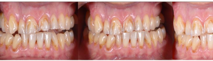
A 68-year-old male patient reported to the clinic with a chief complaint of dislodged implant prosthesis. clinical examination revealed that the mandible was severely resorbed and there was dislodged implant abutment w.r.t. 33 and implant mobility w.r.t. 43 (Figure 1).

Figure 1: Preoperative Intraoral view- Mandibular Arch.
The implants were placed 4 years back. Orthopantomograph and CBCT was advised to evaluate the status of bone availability and implants. It was decided to remove the mobile implant and submerge the other implant, following which new implants will be placed depending on the bone availability. Patient was explained about the treatment plan and he agreed for the same. Thorough medical history was taken along with patient consent.
Diagnostic denture was fabricated using conventional steps. Primary impression was taken using putty and light body (Zetaplus C-silicone, Zhermack, Italy). Custom tray was fabricated on the primary cast. Border moulding was done using low fusing impression compound (DPI pinnacle tracing sticks, DPI, India) and final impression was recorded using light body (Zetaplus, Zhermack, Italy). Jaw relation was recorded, followed by teeth arrangement and processing of dentures after try-in. The mandibular denture was duplicated using clear acrylic and holes were drilled at the site of implants placement which was to be used as a surgical stent.
A full thickness mucoperiosteal flap was raised, the stent was placed and pilot drills were made according to the stent (Figure 2, 3).


Figure 2: Full thickness flap. Figure 3: Drills placed using stent.
Two implants (ifix, India) of 4mm x 12mm were placed and the mobile implant was removed and the other implant was trimmed so as submerge it. Sutures were placed and the patient was recalled after 1 week for suture removal. A healing period of 3 months was allowed after with the implants were loaded. Second stage surgery was done to expose the implants and locator abutments were placed.


Figure 4: Implant Placement. Figure 5: Locators placement.
Holes were drilled in the mandibular denture at the site of locator abutments (Figure 6) to receive female housings. A soft rubber collar was placed around the abutments (Figure 7) to prevent soft tissue injury during polymerisation of acrylic.

Figure 6: Holes drilled in the denture. Figure 7: Metal Housing with gingival sleeve.
The plastic resilient inserts were placed inside the metal housing and the housings were incorporated on the locators. Cold cure acrylic resin (DPI RR cold cure, DPI, India) was mixed and placed into the hollow space on the intaglio surface of the mandibular denture and the denture was positioned inside the patient’s mouth and the patient was instructed to bite in centric occlusion. The material was allowed to set and excess material was polished and trimmed (Figure 8).

Figure 8: Locator Pickup. Figure 9: Denture insertion.
The denture was reoriented in same position (Figure 9). Depending on the usage and requirements of each patient, the retention can be gradually enhanced by switching to higher retention caps. After the prosthesis was delivered, the patient received instructions regarding the recall and aftercare plan. The patient was recalled and was satisfied with the aesthetics and functions of his dentures (Figure 10,11).


Figure 10: Post operative OPG. Figure 11: Post operative smile.
Discussion
Dentists have an excellent chance to enhance their patients' dental health and quality of life using implant-supported overdentures. When compared to a conventional full denture, the chewing efficiency of an overdenture supported by an implant is increased by over 20%. Problems with mandibular dentures, such as loss of stability or retention, decline in function, difficulties with speech, tissue sensitivity, and soft tissue abrasion, are the main reason for a mandibular implant-supported overdenture [2].
The locator system outperformed the ball and bar attachments in terms of clinical outcomes, according to Cakarer et al. A clinical investigation carried out by El-Sheikh et al. provided more evidence for this. They concluded that in situations of mandibular atrophy, the use of two thin bone level implants with locator attachments is predictable and can support an overdenture sufficiently.3 While the denture cap can rotate over the male in all directions, the male remains in static contact with the female socket. This pivoting action provides increased resilience and inhibits dislodgement by accommodating the natural movements during occlusion [4].
The Locator® has a low vertical profile, dual retention, simplicity of insertion and removal, and a special pivoting and self-aligning capability that increases its resilience and tolerance for implant divergency (up to 40°). Because of these design elements, the Locator® quickly rose to prominence as one of the most widely used stud attachments. As of 2010, it was available for about 350 different implants from seven different manufacturers [5].
Compared to those who received mandibular conventional dentures, the group that received the mandibular implant over denture had a noticeably higher quality of life in terms of dental health one year following treatment. The choice of occlusal concept is influenced by the opposing arch condition. For patients with an edentulous maxilla who oppose an implant-supported overdenture, the majority of practitioners advise a balanced occlusion.
Conclusion
A straightforward, affordable, non-invasive, and more retentive locator attachment overdenture treatment approach for an atrophic mandible has been outlined in this article. This procedure takes less clinical time and stops the remnant alveolar ridge from further resorbing. Most importantly, it increases patient satisfaction by providing a sturdy, comfortable prosthesis with improved functionality. The locator attachment was shown to be more beneficial than the ball attachment. This is especially important when the use of traditional attachment systems is restricted by diverging implants and a small interocclusal space. Additionally, unlike ball attachments, it does not require regular repairs.
References
Genesis Scientific Publication is licensed under CC BY-NC-ND 4.0![]()
![]()
![]()
![]()
