Bone Truss Bridge (BTB) Approach for Atrophic Maxilla Rehabilitation
Jimoh Olubanwo Agbaje1 and Henri Diederich2*
1OMFS-IMPATH Research Group, Department of Imaging and Pathology, Faculty of Medicine, Catholic University Leuven, Belgium
2Doctor in dental medicine 114 av de la Faiencerie, L- 1511 Luxembourg
*Corresponding author: Henri Diederich, Doctor in dental medicine 114 av de la Faiencerie, L- 1511 Luxembourg
Citation: Agbaje JO, Diederich H. Bone Truss Bridge (BTB) Approach for Atrophic Maxilla Rehabilitation. Genesis J Dent Rep. 1(1):1-12.
Received: Febraury 02, 2025 | Published: February 12, 2025
Copyright© 2025 genesis pub by Agbaje JO, et al. CC BY-NC-ND 4.0 DEED. This is an open-access article distributed under the terms of the Creative Commons Attribution-Non Commercial-No Derivatives 4.0 International License. This allows others distribute, remix, tweak, and build upon the work, even commercially, as long as they credit the authors for the original creation.
Abstract
The Bone Truss Bridge (BTB) concept represents a novel and promising approach for dental implant rehabilitation in the atrophic maxilla. It offers a minimally invasive alternative to traditional two-piece implants with angulated abutments. Additionally, the milled metal frame with angulated screw channels allows for easy retrievability in case of complications.
The BTB approach demonstrated high patient satisfaction due to reduced treatment time and a less invasive procedure compared to traditional methods.
The presented case studies highlight successful outcomes. Further research, validation, and broader clinical applications will be essential to further establish the role of the BTB approach in the future of implant dentistry.
Keywords
Bone Truss Bridge, rehabilitation, the atrophic maxilla, minimally invasive approach,
Introduction and Problem Statement
Challenge of atrophic maxilla
The primary problem addressed in this study is the significant challenge posed by maxillary atrophy (severe bone loss in the upper jaw) in dental implant rehabilitation. This atrophy is commonly caused by long-term tooth loss (edentulism), trauma, or congenital conditions [1]. The resulting compromised bone volume and density make traditional implant placement difficult or even impossible [2].
Limitations of traditional methods
Traditional solutions such as bone grafting, sinus lifting, and zygomatic implants are often invasive, complex, time-consuming, and associated with high morbidity and donor site complications, especially in elderly patients [3]. Autologous bone augmentation techniques are regarded as the "gold standard" for treating severely atrophic maxillae. However, these procedures are time-consuming, carry a risk of graft loss, and are frequently associated with donor site morbidity [4].
Need for minimally invasive solutions
There is a clear need for minimally invasive, less complex, and more efficient treatment options that can circumvent the drawbacks of traditional methods. Alternative approaches should prioritize structural integrity, long-term stability, and reduced patient morbidity.
The bone truss bridge (BTB) concept
The Bone Truss Bridge (BTB) approach is an advanced technique in dental implantology designed to rehabilitate patients with severe bone loss or complex anatomical challenges. The Bone Truss Bridge (BTB) concept draws inspiration from the Bowstring Truss Bridge. This innovative design was first patented by Squire Whipple in the United States on April 24, 1841, under Patent No. 2064. Whipple's pioneering approach incorporated cast-iron segments combined with wrought iron diagonal ties or braces to maintain the integrity of the arch under uneven loading conditions [5].
The bone truss bridge (BTB) approach in dental implantology
In dental implantology this method utilizes innovative surgical techniques and strategic implant placement to minimize the need for bone grafting, optimize stability, and provide a durable foundation for prosthetic rehabilitation. By anchoring implants in anatomically favorable locations such as the pterygoid plates, nasal floor, and, in some cases, the zygomatic bone, the BTB approach enhances support and load distribution [6].
Key components
The BTB method utilizes strategically placed implants in the following regions:
- Pterygoid Implants: These provide robust cortical anchorage in the pterygomaxillary region, effectively counteract lateral forces and ensure long-term stability [7].
- Trans-Nasal Implants: These thin, one-piece tissue-level implants are placed directly into the cortical bone around the nasal area using a palatal approach, this allows for minimal bone width requirements [8].
- Long Thin Implants (22-26 mm): These implants can be inserted either obliquely or straight to maximize strong anchorage and structural support.
- Anterior Implants: Additional implants are placed in the anterior segment of the maxilla to enhance support and distribution of occlusal loads.
Ideal implant placement strategy
Ideally, eight implants are placed:
- Two implants in the pterygoid region
- Two implants in the pyriform region
- Four implants in the anterior segment of the maxilla
The exact number of implants is dependent on the available bone volume and density, allowing for a customized approach to each patient's anatomy.
Advantages of the BTB Approach
Enhanced Stability and Load Distribution
The strategic placement of implants in regions with high bone density, such as the pterygoid plates and nasal floor, ensures superior stability and optimal load distribution. This approach reduces stress on individual implants, thereby enhancing long-term success [9,10].
Reduced need for bone grafting
By leveraging existing anatomical structures, the BTB approach minimizes the necessity for extensive bone augmentation. This is particularly advantageous for patients with atrophic jaws, as it decreases surgical complexity, cost, and healing time [11,12].
Decreased surgical and recovery time
The BTB approach employs streamlined protocols and efficient implant placement techniques, leading to shorter surgical durations and faster recovery times compared to traditional grafting methods [13, 14].
Versatility in complex cases
This technique is especially beneficial for patients with severe bone loss or complex anatomical conditions where conventional implant methods may be infeasible. It offers a viable alternative to invasive procedures such as sinus lifts or ridge augmentation [15,16].
Enhanced functionality and aesthetics
Precise implant positioning facilitates optimal prosthetic integration, resulting in improved functional outcomes, such as efficient mastication, and enhanced aesthetics for a more natural-looking smile [9, 7].
Cost-effectiveness
By reducing or eliminating the need for additional surgical procedures like grafting, the BTB approach may lower overall treatment costs [6, 13].
Limitations of the BTB Approach
Technical complexity
The BTB approach requires a high level of clinical expertise. Accurate planning, precise surgical execution, and advanced prosthetic integration are critical for optimal outcomes [10, 7].
Limited practitioner availability
Not all clinicians are trained in advanced techniques such as pterygoid or transnasal implant placement, which may limit patient access to this treatment [11].
Potential Complications
While the need for grafting is reduced, implant placement in unconventional locations, such as the pterygoid plates or nasal floor, carries a risk of complications, including sinus perforations or nerve injuries if not performed with precision [ 12, 11].
Higher initial technological costs
The BTB approach relies on advanced diagnostic tools, such as cone-beam computed tomography (CBCT) imaging, CAD/CAM technology, and customized prosthetic solutions, which may increase the initial treatment costs for clinicians [7, 14].
Patient adaptation
Patients may require time to adapt to the biomechanics of a full-arch restoration, particularly in cases with significant atrophy [9].
Challenges in prosthetic design
Achieving a passive fit in prosthetic design can be challenging in complex cases. Any inaccuracies in prosthetic fabrication may compromise long-term outcomes [14, 15].
Clinical considerations
The BTB approach represents a significant advancement in implantology by addressing the limitations of traditional methods and offering viable solutions for complex cases. However, its success is contingent upon meticulous case selection, comprehensive treatment planning, and the clinician's surgical expertise.
Case 1
A 58-year-old female patient underwent maxillary implant placement 10 years ago. In early 2024, she began experiencing complications with the implants, including bridge fractures and detachment, which result in significant discomfort. She sought an alternative treatment approach, as she was unwilling to undergo bone augmentation or a lateral sinus lift.
During the initial consultation, a clinical examination and OPG scan were performed which reveal minimal residual bone volume. Given the poor condition of the existing implants and bridge, all remaining implants and prosthetic components were removed (Figure 1,2).

Figure 1: Situation of the mouth at first appointment.

Figure 2: Removal of
rest of the implants and bridge.
A full thickness mucoperiosteal flap was raised, extending from the left side to the tuberosity region to facilitate pterygoid implant placement. A ROOTT P 3.5/20 mm implant was inserted in the pterygoid region using a pilot drill followed by self-tapping to achieve a final torque of approximately 70 N/cm.
In positions 21, 11, and 13, three ROOTT C 3.0/16 mm compressive implants were placed with palatal entry points to engage the nasal floor, this ensures optimal primary stability. The achieved insertion torque was approximately 50 N/cm. On the right side, an additional pterygoid implant ROOTT P 3.5/20 mm was placed with a final torque of approximately 60 N/cm (Figure 3,4).

Figure 3: Surgical procedure.
The surgical procedure was to do a big flap by beginning on the left side and preparing the end of the tuberosity to put a pterygoid implant.
The pterygoid implant ROOTT P 3.5/20 mm was selected. The insertion was done just by using first a pilot drill and then by self-tapping the implant was inserted and the final torque was around 70 N/cm.

Figure 4: ROOTT Compressive Implant
C 3.0/ 16 mm.
Following implant placement, sutures were placed, and an impression was taken with screwed transfer copings. A silicone putty impression was taken without a tray.
At the second appointment after 5 days a verification jig was trialed, and bite registration was performed to establish the vertical dimension of occlusion (DVO) (Figure 5).

Figure 5: The verification jig.
At the third appointment an esthetic try-in of the bridge framework was done (Figure 6).

Figure 6: Aesthetic try in.
At the fourth appointment 2 weeks after surgery, the metal framework, fabricated by Camex, the milling center (Luxembourg), was evaluated for fit and precision for the final prosthesis construction (Figure 7).

Figure 7: a) Metal frame; b) Plaster cast.
A lightweight metal-resin bridge was fabricated to minimize excessive loading on the implants due to the limited bone volume. Angulated screw channels were incorporated to optimize the fit (Figure 8,9).

Figure 8: Finish metal resin bridge.

Figure 9: Soft tissue
before bridge placement.
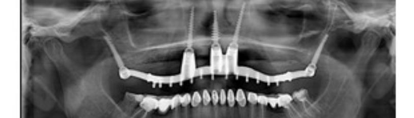
The final bridge was placed and secured three weeks post-surgery (Figure 10,11).

Figure 10: Clinical photo after placement.

Figure 11: OPG after final placement.
Case 2
An 82-year-old male patient presented with missing, fractured, and mobile teeth in the maxilla (Figure 12). He sought a fixed prosthetic solution that was minimally invasive and could be completed within a short timeframe. Following radiological evaluation and treatment plan discussion, the patient opted for a metal-resin bridge due to financial considerations.

Figure 12: OPG at presentation.
At the first appointment teeth the remaining maxillary teeth were extracted, and implants were placed immediately. ROOTT P Compressive Implants were chosen for their high primary stability and potential osseointegration (Figure 13.14). Implant lengths of 20–24 mm was employed to ensure sufficient anchorage.

Figure 13: Tooth extraction and
implant placement.

Figure 14: ROOTT P
Compressive Implants.
The BTB technique was employed, with critical anchorage points including the pterygoid plates and nasal floor region. In certain cases, controlled penetration into the nasal floor was performed to enhance implant stability.
An immediate impression was taken, and a temporary bridge was fabricated at chairside to restore function.
At Second Appointment a verification jig was tried in to confirm implant positioning accuracy. The impression was taken without a tray (Figure 15), and the intraoral validation of the model and jig ensured precision in the final prosthesis fabrication (Figure 16,17).

Figure 15: Impression without tray.

Figure 16: a) Model and b) verification jig.

Figure 17: Try in in the mouth
of the jig.
An esthetic try-in of the future bridge was performed at third appointment to assess fit, occlusion, and appearance. Following validation, the bridge was finalized for definitive placement (Figure18,19).

Figure 18: Aesthetical try in and validation.

Figure 19: Finish metal resin bridge.
Note: There is no reference to Figure 19
At final Appointment, before bridge placement, peri-implant soft tissues were evaluated (Figure 20). On the day of insertion, the metal-resin bridge was secured onto the implants, providing the patient with a functional and esthetic fixed prosthesis (Figure 21,22).

Figure 20: Soft tissue before placement.
The treatment was completed effectively, and successfully achieved the patient's expectations (Figure 21,22).

Figure 21: After bridge placement.

Figure 22: OPG After bridge placement.
Case 3
A 63-year-old male patient presented with severe periodontal problem in the maxilla. His posterior teeth had been lost due to periodontal disease, and the remaining anterior teeth exhibited mobility and suppuration. While periodontal treatment was performed in the mandible, the extent of maxillary damage necessitated complete extraction and immediate rehabilitation (Figure 23,24).

Figure 23: OPG at presentation.
After radiological assessment was conducted to evaluate bone structure and finalize the treatment plan. ROOTT P Compressive implants (TRATE AG) were selected due to their ability to achieve high primary stability in compromised bone conditions.

Figure 24: Clinical photograph
at presentation.
Following the initial assessment, all maxillary teeth were extracted, and implants were subsequently placed. To address the bone defects, grafting material was applied at the extraction sites (Figure 25,26).

Figure 25: Clinical photograph after tooth extraction.

Figure 26: Implant placement and bone defects
filled with bone grafting material. .
Following implant placement and suturing, an immediate impression was taken using screwed transfers. The transfers were stabilized with Luxabite, a rigid occlusal registration material, and a silicone impression was taken without a tray to ensure accuracy (Figure 27-29).

Figure 27: Implant with transfer coping.

Figure 28: Silicon Impression
taken without tray.

Figure 29: Silicon impression
with transfer coping.
Five days post-surgery, a verification jig was tested to confirm implant positioning. Bite registration was performed to ensure accurate occlusal alignment (Figure 30-32).

Figure 30: Verification jig.

Figure 31: Bite plate and wax for bite registration.

Figure 32: Verification jig tried in the mouth.
At the third appointment the metal framework was evaluated for fit and alignment (Figure 33). By this stage, the soft tissues had undergone sufficient healing allowing for optimal adaptation of the prosthesis.

Figure 33: The metal framework.
Ten days later, the final metal-ceramic bridge was fabricated and delivered. Before placement, peri-implant soft tissues were revaluated to confirm adequate healing (fig 34 -.35) The definitive restoration was successfully secured three weeks after initial surgery, providing the patient with a functional and esthetic solution (Figure 36-37).

Figure 34: Metal ceramic bridge.

Figure 35: Soft tissue before placement.

Figure 36: Definitive metal
ceramic bridge after delivery.
The definitive metal ceramic bridge was already delivered after 3 weeks.

Figure 37: OPG after completion of treatment.
This case highlights the efficacy of immediate rehabilitation with ROOTT P implants and a metal-ceramic bridge for patients with advanced periodontal disease. The patient was highly satisfied, as the treatment successfully restored function and aesthetics within a short timeframe.
Technical Details and Materials:
- Implants Used: ROOTT Compressive multi-unit implants (TRATE AG, Switzerland), specifically engineered for narrow ridges and cases of severe atrophy as illustrated in these patient case studies.
- Surgical Procedure: The surgical protocol involves a crestal linear incision, flap elevation, and precise implant placement in the pterygoid, trans-nasal, and anterior regions. The multi-unit design of the implants allows for precise angulation adjustment during the prosthetic phase and supports screw-retained prosthesis fixation.
- Prosthetic Phase: The prosthetic workflow involves obtaining direct impressions with open-tray (with or without tray) impression transfers, followed by the fabrication of a verification jig to ensure a passive fit. The final screw-retained prosthesis is then delivered. Metal frameworks with acrylic or metal-ceramic bridges enable immediate prosthesis placement, enhancing procedural efficiency and patient comfort.
Conclusion
The BTB concept represents a novel and promising approach for dental implant rehabilitation in the atrophic maxilla. It offers a minimally invasive alternative to traditional two-piece implants with angulated abutments. Additionally, the milled metal frame with angulated screw channels allows for retrievability in case of complications.
The BTB approach has demonstrated high patient satisfaction due to reduced treatment time and a less invasive procedure compared to traditional methods.
While the presented case studies highlight successful outcomes, ongoing research, validation, and broader clinical applications will be essential to further establish the role of the BTB approach in the future of implant dentistry.
This document provides an overview of key concepts and findings related to the Bone Truss Bridge (BTB) approach for the rehabilitation of the atrophic maxilla, as described in the available literature. Since this is a relatively new technique, further clinical studies and long-term trials are necessary to fully validate its efficacy and safety.
Reference
Genesis Scientific Publication is licensed under CC BY-NC-ND 4.0![]()
![]()
![]()
![]()
