Aging Management with the Use of Exosomes, Prp/Prf with Albumin Gel (Plasma Gel) and Associated Techniques in Orofacial Harmonization Treatment
Adelaide Maso*
Master in Dental Medicine, Integrative Medicine, Hormonal Pharmacology, Orofacial Harmonization Lecturer
*Corresponding author: Adelaide Maso, Master in Dental Medicine, Integrative Medicine, Hormonal Pharmacology, Orofacial Harmonization Lecturer
Citation: Maso A. (2024) Aging Management with the use of Exosomes, Prp/Prf with Albumin Gel (Plasma Gel) and Associated Techniques in Orofacial Harmonization Treatment. J Stem Cell Res. 5(2):1-9.
Received: June 26, 2024 | Published: July 12, 2024
Copyright© 2024 genesis pub by Maso A. CC BY-NC-ND 4.0 DEED. This is an open-access article distributed under the terms of the Creative Commons Attribution-Non Commercial-No Derivatives 4.0 International License. This allows others distribute, remix, tweak, and build upon the work, even commercially, as long as they credit the authors for the original creation.
DOI: https://doi.org/10.52793/JSCR.2024.5(2)-S2(3)
Abstract
Aging is a physiological process that involves a progressive decline in organ function, with loss of homeostasis and an increased likelihood of illness and death. This account focuses on classical perspectives on exosome biogenesis, and associated age-related changes. Due to their ability to transmit biological information between cells, this work also discusses the interaction of exosomes derived from mesenchymal cells, as a powerful adjunct in the treatment of combining Orofacial Harmonization techniques.
The demand for aesthetic and rejuvenating treatments is increasing in society, therefore, every day, the search for orofacial harmonization increases exponentially with the aim of delaying aging.
Orofacial harmonization is a set of procedures performed by dentists, which aims to achieve aesthetic and functional balance of the face.
Exosomes are membranous extracellular vesicles that range from 30 to 200 nm in diameter. Exosomes have been found to be secreted by most types of cells, including cells of the immune system (B cells, T cells, mast cells, dendritic cells), neuronal cells, epithelial cells, endothelial cells, embryonic cells, cancer cells, and mesenchymal stem cells (MSCs).
The search for youth and perfect skin is a natural human desire. And, along this path, collagen biostimulation stands out as a safe and effective option for rejuvenating the skin, combating the signs of aging and promoting a firmer, more toned and radiant appearance.
Research into exosome therapies continues to thrive. Subsequent data on indications, dose response, safety, efficacy, and ability to combine exosome therapy as a "skin primer" for biostimulation techniques such as calcium hydroxylapatite (CaHA), platelet-rich plasma (PRP), and fibrin matrix platelet-rich plasma membrane (PRFM) is growing rapidly.
Keywords
Exosomes; Facial rejuvenation; Orofacial harmonization; Collagen biostimulator; PRP - Platelet-rich plasma.
Introduction
Facial aging is a multifactorial process that has been widely studied, changes in the skin of the facial skeleton and soft tissues are considered the pillars of aging [1].
The intrinsic process of aging is characterized by a decrease in skin collagen, a decrease in elastin and hyaluronic acid (HA), which generates reduced thickening of the skin and loss of elasticity [2].
The discovery of extracellular vesicles (EVs) or exosomes dates back to the 1940s, and these small vesicles were ignored as cellular garbage disposal sites for a long time [3].
Significant attention came only in the mid-2000s, following the rediscovery of exosomes as messengers of cell-to-cell communication. Exosomes, and specifically MSC-Exos, have great potential for promoting rapid and efficient wound healing. Exosomes can be applied directly to a lesion, which in animal models has been shown to promote collagen synthesis and the proliferation and migration of fibroblasts and keratinocytes. These effects have been shown to be due in part to exosomal regulations of microRNA levels and protease activities [4].
Exosomes are derived from mesenchymal stem cells (MSCs) and can potentially be used as an alternative for cell therapy, wound treatment and aid in angiogenesis [5].
Studies of exosome treatments continue to flourish. Subsequent knowledge surrounding indications, dose response, safety, efficacy, and the ability to combine exosome treatment as a "skin primer" - for bio stimulation modalities such as calcium hydroxylapatite (CaHA), platelet-rich plasma (PRP ) and platelet-rich plasma fibrin matrix (PRFM) is growing rapidly [6].
Aging is a complex and natural biological process that involves several different pathways with genetic and environmental elements, which lead to a decline in physical and mental capabilities over the years. The effects of aging occur in several systems of the body, including the endocrine system, which results in the decline of several hormones, known as endocrinosenescence [7].
The accumulation of stochastic molecular and cellular damage is believed to cause aging. Although there is no precise definition of the exact type of damage responsible for aging-related degeneration, it likely includes mitochondrial dysfunction, high levels of ROS, telomeric attrition, changes in nuclear structure, accumulation of genetic mutations, or damage to DNA, proteins, and membranes [8].
Exosomes are involved in multiple physiological and pathological processes, including cellular senescence. Exosomes mediate cross-signaling and play a critical role in cell-cell communications. Exosomes have evolved as potential biomarkers for aging-related diseases.
Development
The skin
The skin is the largest organ of the human body, provides mechanical protection and plays an important role in thermoregulation, water control and exposure to external aggressions, as it is the largest external organ and is constantly exposed to environmental conditions. Being composed of three interconnected layers, epidermis, dermis and hypodermis [9].
In the skin, the aged appearance is represented by wrinkles and sagging and results from structural changes at the molecular level. Modifications in collagen, the most important protein in connective tissue, were responsible for these anatomical changes [10].
The dynamics of skin metabolism are controlled by automatic hormonal functions, such as hydration. In the skin, hormones are responsible for synthesizing hyaluronic acid and producing collagen. Anti-aging therapies and hormone replacement have been a complementary alternative used by the scientific community for a better result in treatment, according to studies, hormone deficiency at ideal levels has a negative response on both the individual's health and skin [2].
Epidermis
It originates in the embryonic leaflet ectoderm, its main function is covering and protection, creating a selective barrier between the external environment and the adjacent connective tissue. It can be divided into 4 strata or layers:
Horny Layer,
Granular Layer
Spinous Layer
Basal Layer ( LUVIZUTO, 2019).
The outermost layer of the skin, the multi-cell layered epidermis, protects the skin from toxins, bacteria and fluid loss through the stratum corneum. Although the fundamental structure of the skin is the same in all humans, there are significant differences in the epidermal architecture of the face between different genders and ethnicities [9].
Dermis
The dermis is the second main part of the skin, aging changes are affected in this layer, it is a highly elastic tissue resistant to wear, it is located just below the epidermis, it is composed of cellular and acellular elements. It is in this layer that we find 70 to 80% of collagen fibers [9].
The fibroblast is the main cell present in the dermis, which is responsible for the synthesis of components of the extracellular matrix (ECM), such as collagen, elastic and reticular fibers. It plays a fundamental role in tissue regeneration [1].
The dermis is divided into three portions:
Papillary, more external.
Reticular, innermost and Perianexial dermis [1].
Hypodermis
The hyopdermis, the deepest layer of the skin, has a thickness that can vary from a few millimeters to several centimeters, and its thickness is extremely important when analyzing facial aging from a volumetric point of view [9].
Subcutaneous tissue, a mesodermal product, lies just beneath the skin. The hypodermis, which is defined by microscopic anatomy as a subcutaneous fascia, is not part of the skin. Instead, it serves as a link between the skin and other muscles and organs [1].
The subcutaneous tissue is composed of a network of dense connective tissue fibers within it, which connects the muscles of the facial mimic to the skin, it is divided into deep and superficial planes by the muscular aponeurotic system [9].
Microscopically, the hypodermis is composed of adipose tissue arranged in lobes, which serves as thermal insulation and maintains caloric homeostasis [1].
Platelet aggregates: PRP/PRF for skin rejuvenation
Platelet-rich plasma (PRP) is an autologous biological product that involves the injection of activated platelets, which stimulate the release of growth factors, triggering fibroblast proliferation and healing through the formation of new collagen, elastin and extracellular matrices [11].
Platelet-rich fibrin (PRF) is a proposed autologous membrane that, while trapping growth factors, offers advantages in the accumulation of platelets and leukocytes in the host. But its faster resorption properties (2 weeks) are limitations. Recent studies have shown that heating a layer of liquid platelet-poor plasma (PPP) can prolong the reabsorption properties of heated albumin (albumin gel) from 2 weeks to more than 4 months (e-PRF) [12].
Marx defines platelet-rich plasma (PRP) as a volume of autologous plasma that has a concentration of platelets above baseline values. (MARX, 2001).
Platelets have a variety of growth factors, and seven growth factors were described by Marx in 2001. Since then, more than 1500 growth factors and regulatory proteins are responsible for the effects of PRP [13].
When producing platelet gel, centrifugation of autologous venous blood results in a high concentration of platelets in a relatively small volume of plasma, resulting in the formation of platelet-rich plasma. This is a blood component composed of thrombin and calcium resulting in the activation of platelets and the beginning of the coagulation stage, resulting in the formation of platelet gel, rich in growth factors such as PDGF, TGF-beta, EGF and VEFG in addition to IGF3-6.8 [14].
Kolster reports that the mechanism of action occurs due to some platelets being activated during the mechanical effect of centrifugation. The release of growth factors occurs after endogenous or exogenous activation of platelets and has a chemotactic effect, as well as a direct and indirect effect on tissue regeneration. Mononuclear leukocyte fibroblasts and mesenchymal stem cells are attracted to PRP, and their proliferation is stimulated [15].
The literature provides indications that the ideal platelet concentration is approximately 2.5 times higher than baseline values, and the greatest secretion of endogenous hyaluronic acid and type 1 procollagen by skin fibroblasts has been described at a concentration in this range [16].
Exosome
Exosomes are produced through the formation of intracellular multivesicular bodies with intraluminal vesicles and double invagination of the plasma membrane. Exosomes may contain membrane proteins, cytosolic and nuclear proteins, extracellular matrix proteins, metabolites and nucleic acids, namely mRNA, non-coding RNA species and DNA [6].
Exosomes are a type of nanometer-sized extracellular vesicles (EVs) released by almost all eukaryotic cells, which serve as mediators for intercellular communication and can be used as cell-free therapies [17].
Mesenchymal cell (MSC)-derived exosomes play a crucial role in modulating the inflammatory and immune response. Increasing evidence indicates that these exosomes promote the polarization of macrophages from the M1 phenotype, associated with pro-inflammatory responses, to the M2 phenotype, which is characterized by the secretion of anti-inflammatory factors such as IL-10 and TGF-β [18].
Cells which have the capacity for differentiation and self-renewal, are being studied with the aim of finding a clinical application in regenerative medicine. Previous studies have demonstrated that mesenchymal stem cells (MSCs) are capable of self-renewal and multipotential differentiation capacity, which demonstrates promising therapeutic potential for tissue regeneration and recovery of skin function [19].
Exosomes released from different cell types contain different lipids and proteins. The lipid composition of exosomes comprises lipids that are part of the plasma membrane and the Golgi, and are enriched in glycosphingolipids, cholesterol phosphatidylserine and ceramide [20].
Skin wound healing is characterized by the repair of injured tissue, with tissue regeneration orchestrated by multiple cells to reestablish a protective barrier.
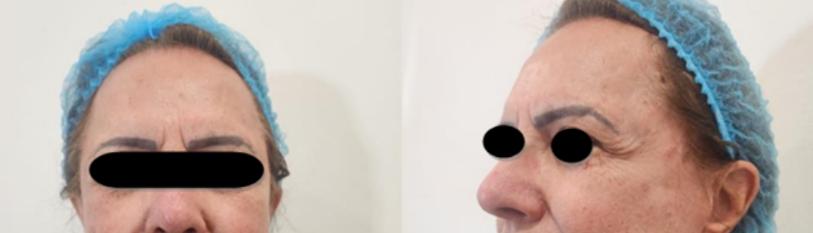
Case Report
Female patient, 78 years old, Caucasian, with wrinkled, sagging, dehydrated and tired-looking skin. He sought Orofacial harmonization treatment, to improve his skin in general. Main complaint wrinkles on the face and “aged and droopy look” Anamnesis, clinical examination, physical examination and evaluation of inflammatory parameters and facial falls were carried out.
On physical examination, flaccid, dehydrated skin was found, with marked wrinkles in the oro-labial and peri-orbital regions.
Treatment
The treatment proposed and carried out on the patient was facial harmonization with several procedures together associated with exosomes. Among them, botulinum toxin, lip filler with hyaluronic acid, skin bio revitalization with exosomes, and collagen bio stimulation with Alb-PRF associated with exosomes.
After anamnesis and clinical examination, the diagnosis was made to begin the procedures. To carry out the procedures, we divide the care steps. Firstly, we performed treatment with botulinum toxin, cleaned the skin with chlorhexidine, and applied it to specific points to relax the muscles.
After the 10th day, biorevitalization of the skin was performed through microneedling using exosomes. Exosomes were applied in two ways, through microneedling and in conjunction with PRF and Alb PRF (plasma gel). To obtain the PRP/PRF, the patient's blood was collected, the total blood collected was from the peripheral region, in 9 mL plastic tubes and centrifuged at 1600 RPM for 5 minutes at a G FORCE 200. Centrifugation by sedimentation compact disc format (CDC) uses relatively low G-force (about 200-500 G). After that, the plasma layer was heated at 80°C for 10 minutes to create denatured albumin (albumin gel). The remaining cells and the growth factor found in the buffy coat (liquid PRF) were then mixed again with the cooled albumin gel to form Alb-PRF, and the same was mixed with already reconstituted exosomes and injected into the patient's face in specific regions using cannulas, thus aiming to improve dermal thickness.
Final Considerations
Many fields of regenerative medicine use platelet concentrates because they can provide supraphysiological concentrations of autologous platelets, leukocytes, and growth factors. To accelerate the formation of hard or soft tissue, regenerative medicine has used a variety of biomaterials, surgical procedures, and growth factors.
Exosomes, and specifically those from mesenchymal cells, have great potential for promoting rapid and efficient wound healing, when associated. Authors report that platelet-rich fibrin (PRF) is a regenerative biomaterial that is completely reabsorbed in approximately 2 to 3 weeks. However, a new heating process has recently been shown to extend the working properties of PRP/PRF from 2 to 3 weeks to 4 to 6 months.
However, many authors report that platelet-rich fibrin (PRF) has been characterized as a regenerative biomaterial that is completely reabsorbed in a typical period of 2 to 3 weeks, but recently, a new heating process has been demonstrated to extend the properties of PRP/PRF work from a standard period of 2 to 3 weeks to a duration of 4 to 6 months.
In order to have a good result and a good response and maintenance of the aesthetic procedures that we want to achieve and maintain for a longer period of time, it is of fundamental importance to manage aging with the use of safe and advanced techniques, guide the patient regarding lifestyle and adequate nutrition and supplementation.
Taking into consideration the articles and findings in this report, we can see the importance of approaching the patient as a whole, and highlighting that exosomes have also gained wide attention in the field of biomarker research and are now seen as a strategy alternative to stem cell-based regenerative therapies.
Although exosomes have achieved significant achievements in several therapies, challenges remain, as we find numerous articles in the literature that report the use of exosomes and their relationship with the skin. However, we need to carry out more studies with this type of treatment (Figure 1).

Figure 1: Clinical Result Before and after.
References
- Luvizoto E, et al. Facial Architecture. 1st ed. São Paulo: Napoleão Quintessence, 2019. 70p.
- Maso A, Tramontini L. (2023) The Benefits of Nanostructured Transdermal Hormonal Replacement Therapy In Orofacial Harmozization Treatment: Case Report. Health Studies. 3(3):103-24.
- Théry C, Zitvogel L, Amigorena S. (2002) Exosomes: Composition, biogenesis and function. Nat Rev. 2(8):569–79.
- Hade MD, Suire CN, Suo Z. (2021) Mesenchymal stem cellderived exosomes: applications in regenerative medicine. Cells 10(8):1959.
- Saheera S, Potnuri AG, Krishnamurthy P. (2020) Nano-Vesicle (Mis)Communication in Senescence-Related Pathologies. Cells. 9(9):1974.
- Kalluri R, LeBleu VS. (2020) The biology, function, and biomedical applications of exosomes. Science. 367(6478):eaau6977.
- Xu D, Tahara H. (2013) The role of exosomes and microRNAs in senescence and aging. Adv Droga Deliv Rev. 65(3):368-75.
- Robbins PD. (2017) Extracellular vesicles and aging. Stem Cell Research. 19(4):98.
- Perlingero A. Sculpting Faces Science & Art in Orofacial Harmonization. 1st ed.São Paulo:Napoleão, 2020:89-91
- Faria JC, Tuma Júnior P, Costa MP, Quagliano AP, Ferreira MC. (1995) Skin aging and collagen. Rev Hosp Clin Fac Med Sao Paulo. 50 Suppl:39-43.
- Phoebe LKW, Lee KWA, Chan LKW, Hung LC, Wu R, et al. (2024) Use of platelet rich plasma for skin rejuvenation. Skin Res Technol. 30(4):e13714..
- Fujioka-Kobayashi M, Schaller B, Mourão CFDAB, Zhang Y, Sculean A, et al. (2020) Biological characterization of an injectable platelet-rich fibrin mixture composed of autologous albumin gel and platelet-rich liquid fibrin (Alb-PRF). Platelets. 32(1):74–81.
- Marx RE. (2001) Platelet Rich Plasma (PRP) What is PRP and what is not PRP? Implant Dent 10(4):225-28
- Eppley BL, Woodell JE, Higgins J. (2004) Platelet quantification and growth factor analysis from platelet-rich plasma: implications for wound healing. Plast Reconstr Surg. 114(6):1502-08.
- https://www.quintessence-publishing.com/usa/en/product/illustrated-guide-to-collagen-induction-with-platelet-rich-plasma-prp
- Anitua E, Sanchez M, Zalduendo MM, et al, (2009). Fibroblastic response to treatment with different preparations rich in growth factors. Cell Prolif 42(2):162-70
- Ding J, Wang X, Chen B, Zhang J, Xu J. (2019) Deferoxamine-stimulated human bone marrow mesenchymal stem cell-derived exosomes accelerate skin wound healing by promoting angiogenesis. BioMed Res Int. 2019:9742765.
- Cunnane EM, Weinbaum JS, O'Brien FJ, Dorp DA. (2018) Future perspective on the role of stem cells and extracellular vesicles in vascular tissue regeneration. Front Cardiovasc Med. 5:86.
- McFarlin K, Gao X, Liu YB, Dulchavsky DS, Kwon D, et al. (2006) Wound Repair Regen. 14(4):471–78.
- Amigorena S. (1998) Antitumor immunotherapy using dendritic cell-derived exosomes. Immunology Research. 149(7-8):661-62.
This article was originally published in a special issue entitled “Precision Cell Therapy”, handled by Editor Dr. Roni Moya.
Genesis Scientific Publication is licensed under CC BY-NC-ND 4.0![]()
![]()
![]()
![]()
