A Case Study on Anesthesia for Percutaneous Closure of Large Secundum ASD in a Patient with Nemaline Myopathy Type
Sara Abou Al-Saud1*, Hossam Walley2, Hatem Desoki2, and Zaki Alzaher2
1 Cardiac Sciences Department, College of Medicine, King Saud University
2 Anesthesia Department in Saud Albabtin Cardiac Center, KSA
*Corresponding author: Sara Abou Al-Saud, Cardiac Sciences Department, College of Medicine, King Saud University
Citation: Al-Saud SA, Walley H, Desoki H and Alzaher Z. (2024) Ilioinguinal/Iliohypogsstric Nerve Block Vs General Anesthesia in Varicocelectomy Surgery. Adv Clin Med Res. 5(4):1-08.
Received: August 22, 2024 | Published: September 10, 2024
Copyright© 2024 genesis pub by AL-Saud SA, et al. CC BY-NC-ND 4.0 DEED. This is an open-access article distributedunder the terms of the Creative Commons Attribution-NonCommercial-No Derivatives 4.0 International License.,This allows others distribute, remix, tweak, and build upon the work, even commercially, as long as they credit the authors for the original creation.
Abstract
Nemaline myopathy (NM) is a rare condition affecting the skeletal muscles and is associated with one in 50,000 live births. Genetic mutations cause this disorder, with the nebulin gene (NEB) being the primary cause of NM. The mutations occur sporadically or can be inherited through autosomal recessive or dominant patterns. NM symptoms vary with severity, including motor problems, body weakness, scoliosis, hypotonia, breathing difficulties and respiratory infections. This case study investigates a 5-year-old girl diagnosed with an atrial septal defect (ASD) and other conditions associated with NM. Thorough family history, genetic and blood testing, she was appropriately diagnosed with nemaline myopathy type 2, represented by a case of ASD.
This case study examines anaesthesia's effect in treating ASD in a rare nemaline myopathy type 2 (NEM2) case. The comorbidities led to using different medications in the percutaneous closure of the ASD by a device. Fentanyl and ketamine were the general anaesthesia used. Rocuronium muscle relaxant facilitated endotracheal intubation, while Sevoflurane maintained the anaesthesia levels for the procedure. At the end of the procedure, Sugammadex muscle relaxant facilitated smooth extubation. Careful selection of medication ensured the patient was hemodynamically stable throughout the procedure.
Keywords
Congenital heart diseases; Atrial septical defect; Nemaline myopathy; Nebulin
Introduction
Nemaline myopathy (NM), also known as congenital rod disease or rod-body myopathy, is a rare neuromuscular condition representing collective muscle disorders [1] The disorder is very common in childhood and at birth, impacting roughly one in every 50,000 live births [2]. Six NM subtypes are categorized based on severity including severe congenital, Amish, intermediate congenital, typical congenital, childhood-onset, and adult-onset [3]. Being rare, there are few resources and little research on NM; therefore, it becomes difficult to diagnose and control it. Severe congenital NM presents at birth with severe muscle weakness and respiratory complications and is usually associated with poor prognosis [3]. Whereas the Amish NM is unique to the Amish population and is characterized by early onset, slow progression, and respiratory involvement. The most common form of NM is the typical congenital and is characterized by generalized muscle weakness and the presence of nemaline bodies in muscle tissue [3]. Those Nemaline bodies consist of accumulations of muscle proteins due to mutations in genes which encode proteins components of the muscle thin filament [4]. Intermediate congenital NM shows symptoms between the severe congenital and typical forms, with moderate muscle weakness and a variable age of onset [3]. Depending on the onset of NM; adult-onset NM manifests in adulthood with progressive muscle weakness and respiratory problems, whereas, child-onset NM appears around 10 years of age and is characterized by distal muscle weakness, and sometimes slowness of muscle contraction [3].
Mutations of the genes encoding proteins in skeletal muscle fibre cells cause NM [4]. A dilemma on the factors behind the mutations holds genetic and environmental factors responsible. Sixty-three per cent of affected children exhibit sporadic mutations, while the remaining 37% follow genetic heredity, where mutated genes are inherited mostly in an autosomal recessive form (24%) and less likely in an autosomal dominant pattern (13%) [4]. NM is caused by mutations in several genes, including NEB, ACTA1, TPM3, TPM2, TNNT1, CFL2, KBTBD13, MYPN, KLHL40, LMOD3, MYO18B, and KLHL41, among others [1]. The most common gene mutations are found in the NEB (50%) and α-actin genes (25%-15%), while the least frequent are TPM3 and TPM2 mutations [3].
Histologically, some genes encode proteins in sarcomeres that are involved in muscle structure and function, whereas other genes act as degradation regulators of the thin actin filaments in muscles [5]. Gene mutations cause small rod-like structures to form in the muscle fibers, originating from the Z-disk [6]. These mutations disrupt the organization of Z-disk proteins, impairing muscle contraction by altering the filament structure or its turnover rate [3]. The mutations' structure continually grows with actin filaments from the Z-disk and consists of α-actinin, NEB proteins, and desmin [6]. Genes carry instructions for producing proteins necessary in skeletal muscles; therefore, their mutations alter the instructions and, thus, the functioning of skeletal muscles [3].
The mutated rod-like structures permit the conduction of muscle biopsies from which NM can be diagnosed. When examined using the Gomori trichrome method, nemaline rods appear as red spots [1]. Electron microscopy is used to distinguish them from mitochondria, which also appear red. Additionally, blood tests are performed to measure elevated creatine kinase levels, a muscle enzyme linked to NM [7]. Therefore, genetic testing to examine genes associated with NM is beneficial for individuals with a family history of the condition. Other methods used to diagnose NM include ultrasound, a physical exam, magnetic resonance imaging, and muscle strength tests [8].
NM victims undergo mild to very severe symptoms, the most common being body weakness [1]. The genetic mutation causes a deformed muscle structure, which leads to weakness, especially of the proximal and facial muscles [3]. NM is linked to other conditions such as hypotonia, respiratory infections, joint contractures, scoliosis, cardiomyopathy and sleep apnea [1]. The sub-types of NM manifest the stated symptoms on different severity levels. For example, the severe congenital type is associated with profound breathing difficulties, swallowing issues, significant muscle weakness and floppiness, and a shortened lifespan [9]. The mild and typical congenital forms possess almost similar symptoms, including respiratory problems and delays in motor milestones [9,10].
No cure or therapy was invented for the treatment of NM [8]. If symptoms are not diagnosed and treated early, the disease will continue to progress. Symptoms present dictate the treatment procedure for the patients. Patients with mild symptoms are prescribed medications to improve skeletal muscle function [9], but surgical treatment is preferred for conditions such as facial deformities and scoliosis [4] management in surgery necessitate the careful selection of medications. Successful medications and anaesthetic techniques include sedation, inhalation anaesthetics, neuraxial technique, logical-regional and total intravenous anaesthetic [4]. This study analyses a nemaline myopathy type 2 (NEM2) case with ASD.
Case Presentation
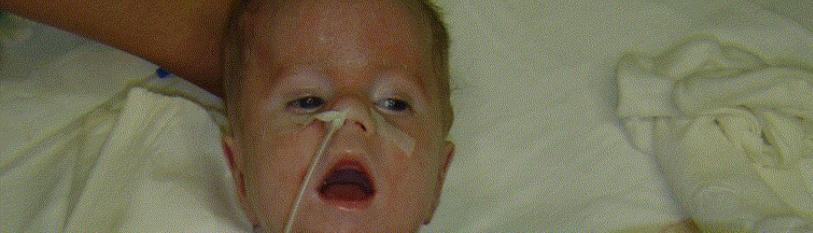
A five-year-old female child was diagnosed with atrial septal defect (ASD), exercise intolerance, muscle weakness, easy fatigability, mild respiratory insufficiency, and muscular hypotonia in infancy. There is evidence that his parents were related by blood and both died during his childhood. Two brothers died in adolescence, and one sister in childhood-onset. The patient was clinically suspected of having an autosomal recessive disorder which accounts for the congenital anomalies. The prior patient examination ruled out spinal muscular atrophy with the deletion/duplication analysis of survival motor neuron 1 (SMN1) gene resulting in negative results. Further diagnosis through genetic and blood testing was conducted to establish facts.
Genetic Testing
A congenital myopathy panel tested a variety of pathogenic gene variants based on their casualty and pathogenicity. CENTOGENE data on gene mutations demonstrates test quality. Targeted genes in the test were ACTA1, NEB, CCDC78, CFL2, BIN1, DNM2, MTM1, MYH7, MTMR14, FHL1, MAMLD1, TNNTI, KBTBD13, MYF6, TPM3, TPM2. The patient was identified with a heterozygous NEB gene variant of uncertain significance. The NEB gene is associated with autosomal recessive NEM2. The patient was hypothesized to have a positive family history of NM; thus, genetic counselling was recommended. No other clinically relevant gene variants were identified.
Blood Testing
Blood drawn from the patient was screened in the lab. The patient had an O+ve blood group, and the levels of the blood components were measured including; haemoglobin level (Hb), white blood cells level (WBC), platelets, prothrombin time (PT), urea, creatine, partial thromboplastin time (PTT), sodium (Na), potassium (K), chloride (Cl), bilirubin, albumin, and the aspartate transferase content (AST) as shown in Table 1. From the blood test results, sickle cell anaemia was ruled out as a condition affecting the patient.
|
Lab Tests |
Results |
Reference Range |
|
Hb |
13.5 g/dL |
12-16 g/dL |
|
WBC |
5.61 x 10⁹/L |
4.5-11.0 x 10⁹/L |
|
Platelets count |
296,000 / μL |
150,000-450,000 / μL |
|
PT |
12.7 seconds |
12.3-15.1 seconds |
|
PTT |
34.6 seconds |
60-70 seconds |
|
Urea |
33.8 mg/dL |
5-20 mg/dL |
|
Creatinine |
0.5 mg/dL |
0.7-1.3 mg/dL |
|
Na |
142 mmol/L |
136-145 mmol/L |
|
K |
4.4 mmol/L |
3.7-5.2 mEq/L |
|
Cl |
107 mEq/L |
96-106 mEq/L |
|
Bilirubin |
0.46 mg/dL |
0.1-1.2 mg/dL |
|
Albumin |
4.2 g/dL |
3.4-5.4 g/dL |
|
AST |
31IU/L |
8.00-48.00 IU/L |
Table 1: Blood testing results.
A multidisciplinary team consisting of pediatric cardiologist, pediatric cardio-thoracic surgeon, interventional pediatric cardiologist, surgeon and cardiac anesthesiologist had revised the transthoracic and transesophageal echo, lab investigations and other associated comorbidities. Because the anatomy of the ASD is amenable to percutaneous closure and considering the hereditary myopathy, a minimally invasive procedure is recommended to avoid delayed recovery and the need for recuperation for postoperative mechanical ventilation, the decision was toward percutaneous closure of the ASD by the ASD device closure that was used as per manufacturer’s instructions (Amplatzer Septal Occluder, Abbott). The device's principle is rooted in the conjoint waist, which acts as a stent to both fixate and occlude ASD. Constructed from a nitinol wire mesh, it consists of a left atrial retention disc, a self-centering stent, and a right atrial disc. A central screw at the proximal end of the device attaches it to the delivery cable. Prior to insertion, the device is collapsed into a tubular loader by pulling on the delivery wire. A 6F delivery sheath is utilized, tailored to fit the device's size.
Risk stratification and suspected complications regarding the procedure, general anaesthesia, postoperative recovery and alternatives were explained clearly and discussed with the family by a cardiologist and cardiac anesthesiologist in the clinic, and informed consent was obtained. Preoperative preparation was done by obtaining informed consent and checking the availability of blood products and postoperative intensive care unit bed. Preoperative premedication was omitted and replaced on the day before the procedure due to hypotonia associated with NM following the child and family reassurance.
On the day of the procedure, the patient was received in the cath lab preparation room and sedated with an intravenous injection of 10mg of Ketamine (0.5mg/Kg, Hikma Farmaceuticals) and shifted to the procedure room in the cath lab. American Society of anesthesiologists standard monitoring was applied to the patient, and general anaesthesia was conducted smoothly and uneventfully by adding 10mg of Ketamine (0.5mg/Kg) and 25mics of Fentanyl (1mic/Kg, Fresenius Kabi). A non-depolarising muscle relaxant facilitated endotracheal intubation with 15mg of Rocuronium (0.7mg/Kg, Hikma Farmaceuticals). Intubation was done with no difficulties by direct laryngoscope with cuffed endotracheal tube size five, and ventilation started with volume-controlled mode on FiO2 of 0.5. The maintenance of anaesthesia was achieved by inhalational anaesthetic in the form of Sevoflurane. Consequently, transesophageal echocardiography and an oral temperature probe were inserted smoothly after which an additional dose of muscle relaxant was given. The patient was hemodynamically stable throughout the procedure, which was completed within 90 minutes.
A nerve stimulator (train-of-four monitor) assessed motor power throughout the procedure. Muscle relaxation was reversed to facilitate smooth extubation by injecting Sugammadex at the end of the procedure. The postoperative Alderet score was 10 points at first 15 minutes, and the patient was discharged to a ward after 1 hour in the recovery area.
Discussion
Most ASDs may be evident among genetic disorder symptoms, as observed in the five-year-old girl's NM case because of their hereditary tendency associated with genetic disorders [11]. Studies hypothesize that an autosomal recessive pattern is ideal to define the inheritance of congenital heart disease, with cases from consanguineous couples leading to an increased risk of congenital heart disease [12]. NM disorders are characterized by nemaline structures (bodies or rods) in skeletal muscle fibers [13]. The disorder is evident at birth, during childhood stages as depicted in the patient and her family, and most rarely in adulthood [14]. Because nemaline rods affect skeletal muscles, NM victims primarily experience weakness throughout the body, especially in the face, trunk, neck and proximal muscles. Progressive weakness can lead to severe cases of facial dysmorphism, difficulty swallowing, skeletal deformities (foot deformities, scoliosis, joint deformities), breathing difficulties, respiratory infections and movement disorders [15]. Common symptoms of NM and ASD are muscle weakness, breathing problems, and easy fatigability [15,16]. NM and ASD existence in the patient's body causes an overlap of symptoms that require creative and effective management.
At least 12 different gene mutations cause NM, with the prominent genes being NEB and α-actin [13]. Various gene mutations form the basis for the spectrum of NM, ranging from NM type 1 to NM type 10 (NEM1-NEM10). Mutations in α-tropomyosin cause NEM1, nebulin, NEM2, α-actin, NEM3, β-tropomyosin, NEM5, KBTBD13, NEM7, KELCH-like 40, NEM7, etc. 41 for NEM9 and leiomodin 3 for NEM10 [17]. With prominent NEB and α-actin gene mutations, NEM2 and NEM3 become the most common NM cases.
Each type of NEM stated has different levels of severity. Considering NEM2 diagnosed in the case study, the disorder can be presented in different severity classes, with the most clinical being early onset [18]. According to Medline, there are six types of NM based on severity; severe congenital, Amish, typical congenital, adult-onset, and childhood-onset [15]. The most common type of the disease is the typical congenital type, and the most lethal is the severe congenital type [15]. Moreover, most victims don't make it past early childhood in the severe NM type due to respiratory difficulties. It is difficult to establish a connection between the level of symptoms' severity and the type of gene involved [19]. However, some connections have unfolded. For instance, TNNT1 is evident in the Amish population to cause severe symptoms at a young age [19].
NEM2 is classified in the typical congenital NM form based on the symptoms, hypotonia and mild respiratory difficulties [19]. Additional symptoms in this category include feeding difficulties and muscle weakness around the trunk, which appears to progress to more distal muscles [19]. NM is a progressive disorder, and in its more severe forms, it leads to pronounced muscle weakness and floppiness, significant breathing difficulties, problems with sucking and swallowing, and limited movement. In most cases, death occurs early if not managed [19]. NM symptoms can be fatal when combined with heart disease. Therefore, early detection of cardiac diseases is vital for the easy management of NM. Cardiac issues in NM patients can lead to heart failure if respiratory muscle weakness develops [20]. Failure to close ASDs in childhood-onset would lead to increased risks of other infections at an older age, such as stroke, pulmonary hypertension, and arrhythmia [11]. Early control of ASD is therefore necessary to parallel ongoing NM treatment for the child-patient.
Currently, there is no cure for NM [21]. However, ASD can be cured through heart surgery and catheterization or may require no treatment for small ASDs [11]. The treatment depends on the severity of the defect, location in the heart, size and age of the child [11]. To control ASD in the presence of NM, management of NM symptoms is essential, and this is done through methods aimed at increasing the mobility and function of the skeletal muscles. NM control methods include medication for better muscle functioning and strength, respiratory support and gastronomy in severe cases, night-time ventilation, physical exercise, and physiotherapy [22]. Antibiotics are also administered, particularly for conditions related to breathing complications. New therapies, such as pharmacological approaches and gene therapy, are being evaluated and developed for the condition [23].
Conclusion
CHDs are susceptible diseases that occur in collaboration with congenital myopathies such as NM on rare occasions like this case. Genetic counselling is necessary for couples before and after having children since offspring can inherit NM. Early diagnosis of symptoms is required for sporadic cases to control CHDs and NM before the defects progress to severe cases.
Key Clinical Message
The anesthetic management of patients with NM should be deliberate and individualized. "Close attention should be given to airway and cardiopulmonary status in this population. These patients should have optimization of their pulmonary function and treatment of any pulmonary infection prior to elective surgery. Currently there are no contraindicated anesthetic techniques, and muscle relaxants can be used. The choice of paralytic agents depends upon the type of surgery and the feasibility of implementing it. As vascular access and airway access may be difficult, the assistance of another anesthesiologist may be helpful. Paying attention to padding and positioning in long and complex surgeries to minimize neuropathy is important. Lastly, patients with NM may benefit from the intensive care unit postoperatively.
References
- Sewry CA, Laitila JM, Wallgren-Pettersson C. (2019) Nemaline myopathies: a current view. Journal of Muscle Res Cell Motil. 40(2):111–26.
- Christophers B, Lopez MA, Gupta VA, Vogel H, Baylies M. (2022) Pediatric Nemaline Myopathy: A Systematic Review Using Individual Patient Data. J Child Neurol. 37(7):652–63.
- https://medlineplus.gov/genetics/condition/nemaline-myopathy/
- Smith T. (2017) Anesthetic consideration for patients with nemaline rod myopathy: a literature review. Pediat Anes and Critical Care J. 5(1):31–9.
- https://www.sciencedirect.com/science/article/pii/B9780124170445000287
- https://www.sciencedirect.com/topics/medicine-and-dentistry/nemaline-myopathy
- Yin X, Pu CQ, Wang Q, Liu JX, Mao YL. (2014) Clinical and pathological features of patients with nemaline myopathy. Molecular Medicine Reports. 10(1):175–82.
- https://www.cedars-sinai.org/health-library/diseases-and-conditions/m/myopathy.html#:~:text=Physical%20therapy%2C%20supportive%20devices%20such
- https://www.musculardystrophyuk.org/conditions/nemaline-myopathy/symptoms
- https://www.childrenshospital.org/research/labs/beggs-laboratory-research/science/myopathies/nemaline-myopathy
- https://kidshealth.org/en/parents/asd.html#:~:text=If%20there
- Suluba E, Shuwei L, Xia Q, Mwanga A. Congenital heart diseases: genetics, non-inherited risk factors, and signaling pathways. Egypt J Med Human Genet. 21(1).
- Sewry CA, Laitila JM, Wallgren-Pettersson C. (2019) Nemaline myopathies: a current view. J Mus Res and Cell Motility. 40(2):111–26.
- https://my.clevelandclinic.org/health/diseases/24180-nemaline-myopathy
- https://www.sciencedirect.com/topics/medicine-and-dentistry/nemaline-myopathy
- https://www.mayoclinic.org/diseases-conditions/atrial-septal-defect/symptoms-causes/syc-20369715
- https://www.sciencedirect.com/science/article/pii/B9780323033541500900
- https://www.omim.org/entry/256030
- https://www.musculardystrophyuk.org/conditions/nemaline-myopathy/symptoms
- Finsterer J, Stöllberger C. (2015) Review of Cardiac Disease in Nemaline Myopathy. Pediat Neurol. 53(6):473–7.
- Yin X, Pu CQ, Wang Q, Liu JX, Mao YL. (2014) Clinical and pathological features of patients with nemaline myopathy. Mol Med Rep. 10(1):175–82.
- https://www.sciencedirect.com/science/article/pii/B9780124170445000287
- Matsson H, Eason J, Bookwalter CS, Klar J, Gustavsson P, et al. (2007) Alpha-cardiac actin mutations produce atrial septal defects. Human Molec Gene. 17(2):256–65.
Genesis Scientific Publication is licensed under CC BY-NC-ND 4.0![]()
![]()
![]()
![]()
